PILOCYTIC
ASTROCYTOMA ....Text.......Neuropathology
- cases....
For undergraduates:
Neuropathology.
Neuroimaging. |
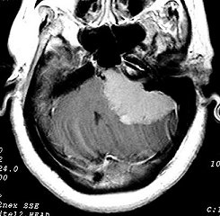
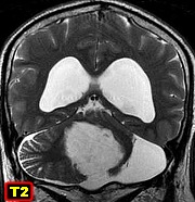
| Cerebellum
- classical cases |
 |
 |
 |
 |
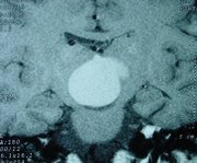
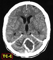
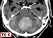
| F. 8 yr.
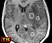
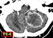
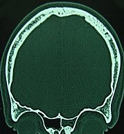
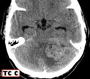
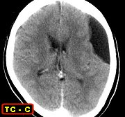
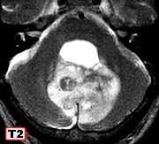
Pilocytic astrocytoma of cerebellum. CT |
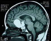
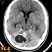
M. 21 yr.
Pilocytic astrocytoma of cerebellum. CT |
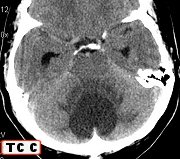
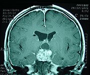
M.
yr. Pilocytic astrocytoma of cerebellum. CT |
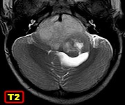
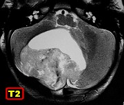
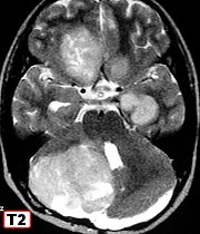
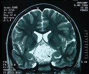
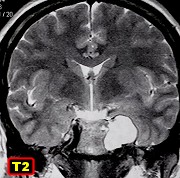
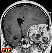
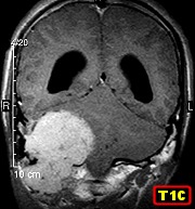
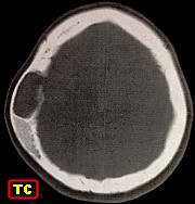
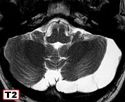
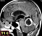
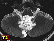
F. 20 yr.
Pilocytic astrocytoma of vermis |
|
|
|
|
 |
 |
 |
 |
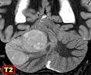
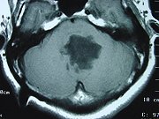
| M. 3 yr.
Pilocytic astrocytoma of vermis |
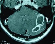
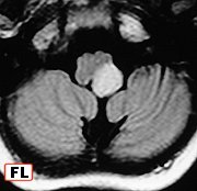
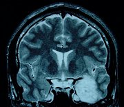
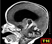
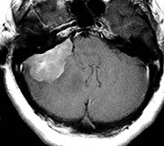
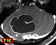
F. 5 yr.
Pilocytic astrocytoma of cerebellar hemisphere |
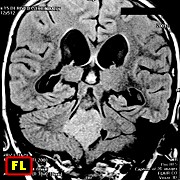
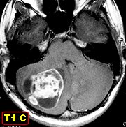
M. 14 yr.
Pilocytic astrocytoma of cerebellar hemisphere |
F. 23 yr.
Pilocytic astrocytoma of cerebellar hemisphere. 17-year follow-up |
|
|
|
|
| Cerebellum
- atypical cases |
 |
 |
 |
 |
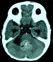
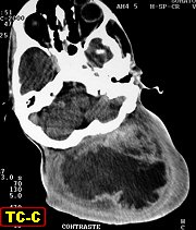
| M. 73 yr.
Pilocytic astrocytoma of vermis in the 8th decade. |
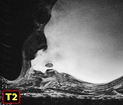
M. 72 yr.
Infiltrating cystic pilocytic astrocytoma of vermis in the 8th decade. |
F. 37 yr.
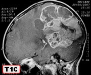
Aggressive pilocytic astrocytoma of cerebellum with extensive meningeal
infiltration |
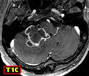
F. 15 yr.
Bilobed, solid and cystic pilocytic astrocytoma of cerebellum with abundant
vascular proliferation |
|
|
|
|
 |
 |
 |
 |
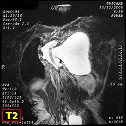
| M.
58 yr. Pilocytic astrocytoma of cerebellar vermis with ventricular seeding.
3-year follow-up |
M. 63 yr.
Cerebellar pilocytic astrocytoma with atypia and high Ki-67 index |
|
|
|
|
 |
 |
|
|
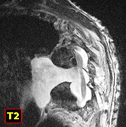
| F. 66 yr.
Pilocytic astrocytoma of cerebellar vermis in the 7th decade |
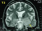
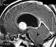
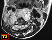
M. 51 yr.
Pilocytic astrocytoma of cerebellum with unusually luxuriant capillary
proliferation (prompting differential diagnosis with hemangioblastoma) |
|
|
|
|
|
|
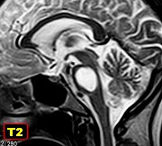
| Midbrain
/ Quadrigeminal plate |
 |
 |
 |
|
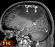
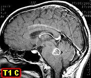
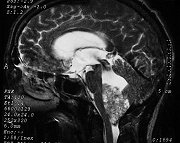
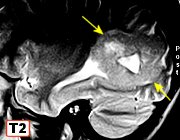
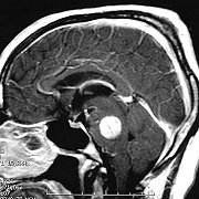
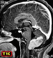
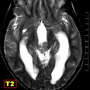
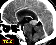
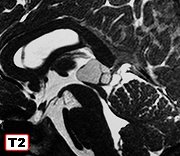
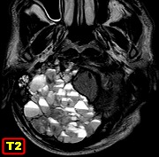
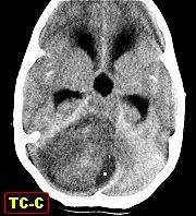
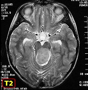
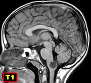
| F. 9 yr.
Pilocytic astrocytoma of quadrigeminal plate |
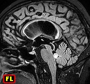
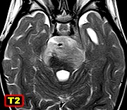
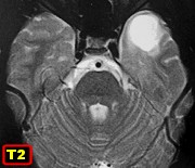
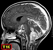
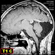
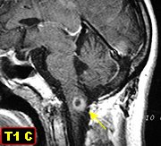
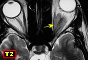
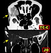
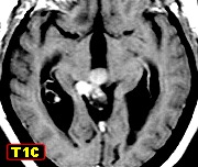
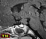
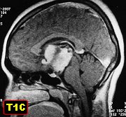
F. 20 yr.
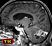
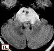
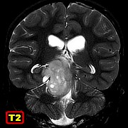
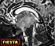
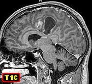
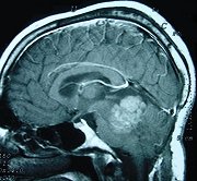
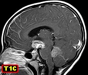
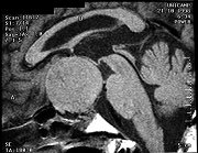
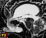
Pilocytic astrocytoma of midbrain |
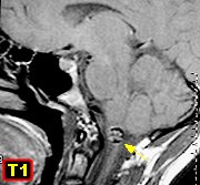
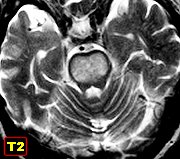
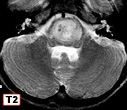
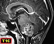
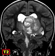
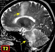
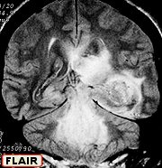
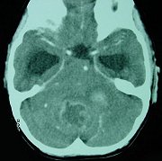
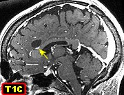
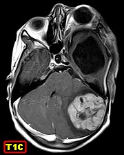
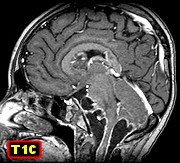
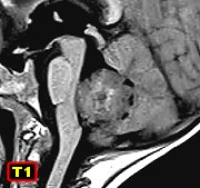
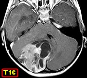
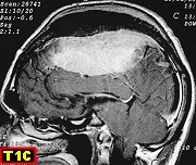
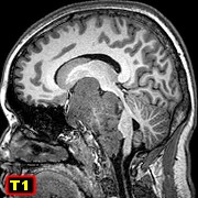
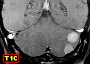
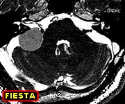
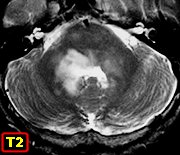
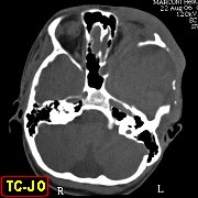
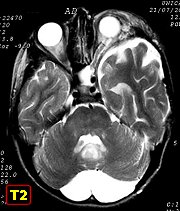
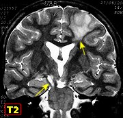
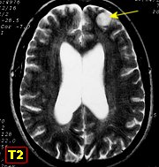
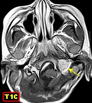
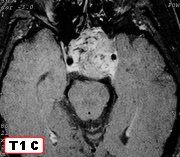
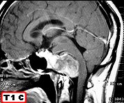
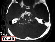
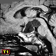
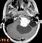
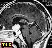
M.
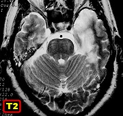
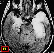
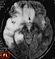
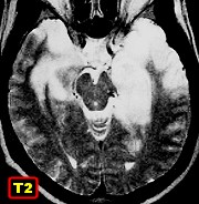
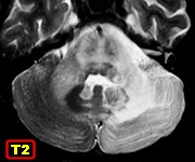
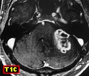
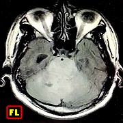
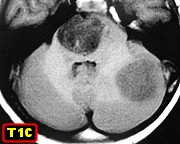
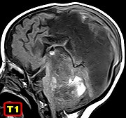
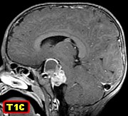
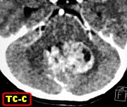
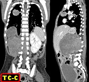
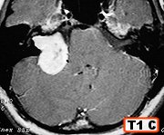
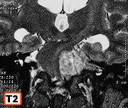
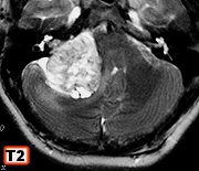
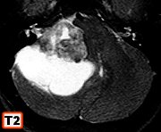
68 yr. Pilocytic astrocytoma of midbrain tegmentum and pons with cerebellar
extension |
|
|
|
|
|
| Midbrain
/ Quadrigeminal plate |
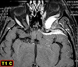
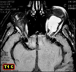
Optic
nerve |
 |
 |
 |
 |
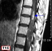
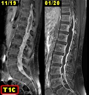
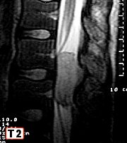
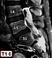
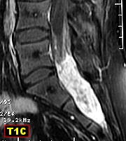
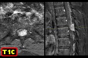
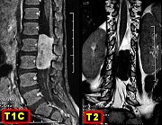
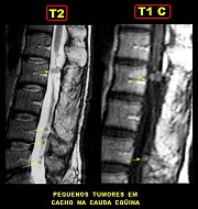
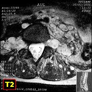
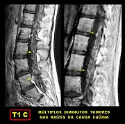
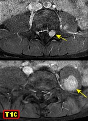
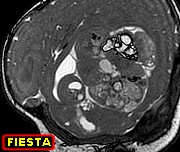
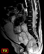
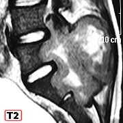
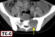
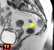
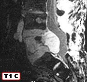
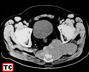
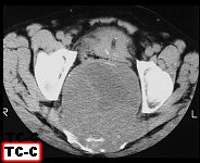
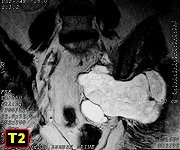
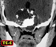
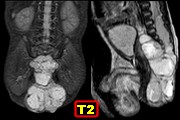
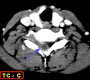
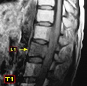
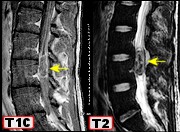
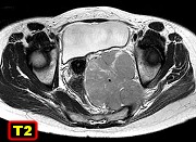
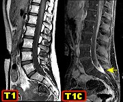
| F.
14 yr. Pilocytic astrocytoma of quadrigeminal plate with CSF
seeding of lumbosacral cul-de-sac |
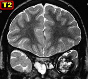
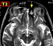
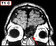
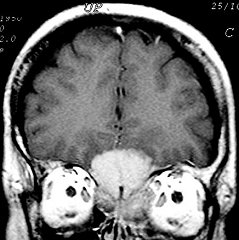
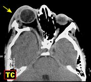
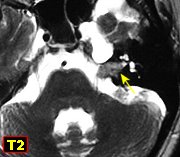
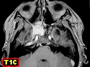
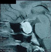
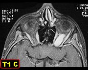
F. 9 yr.
Pilocytic astrocytoma of optic nerve |
M. 11 yr.
Pilocytic astrocytoma of optic nerve. Extension to optic chiasm |
|
|
|
|
| Retina |
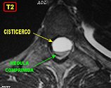
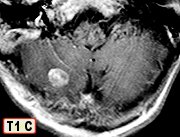
Medulla |
|
|
 |
 |
|
|
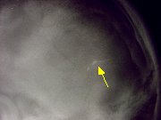
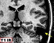
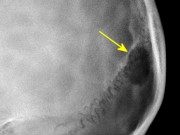
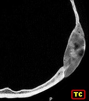
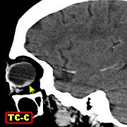
| F. 67 yr.
Pilocytic astrocytoma of retina. Text |
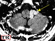
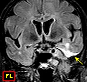
F. 8 yr
7 m. Pilocytic astrocytoma of medulla in NF1 |
|
|
|
|
|
|
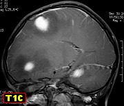
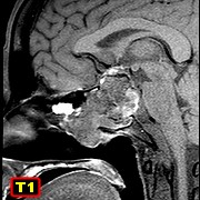
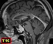
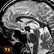
| Hypothalamus,
diencephalon, basal region of cerebral hemispheres |
 |
 |
 |
 |
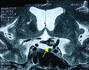
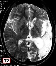
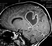
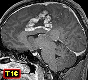
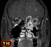
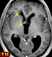
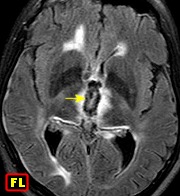
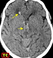
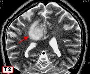
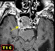
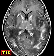
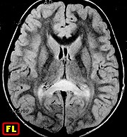
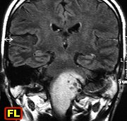
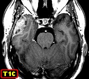
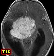
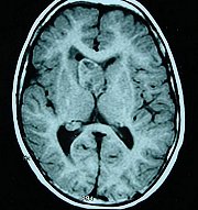
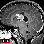
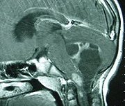
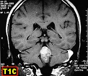
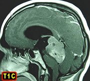
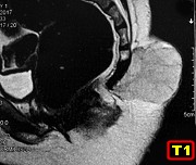
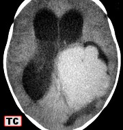
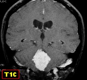
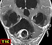
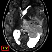
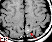
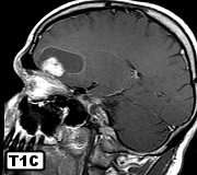
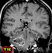
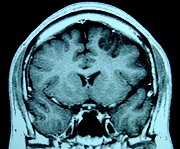
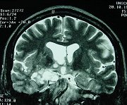
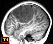
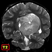
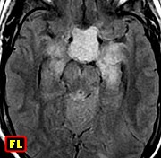
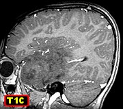
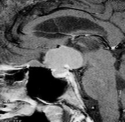
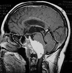
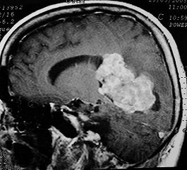
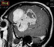
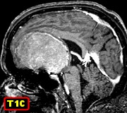
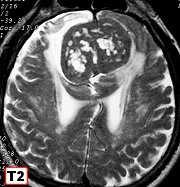
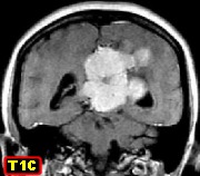
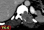
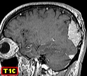
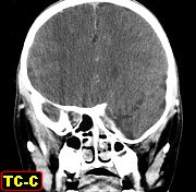
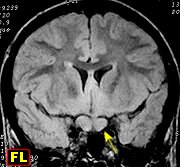
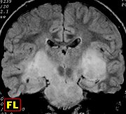
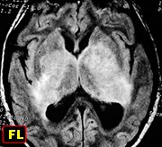
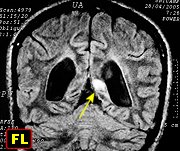
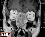
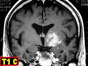
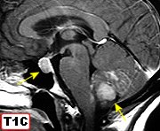
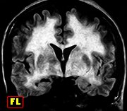
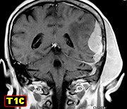
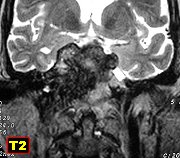
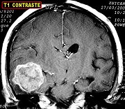
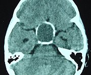
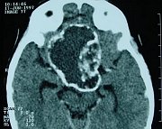
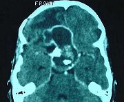
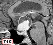
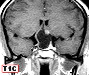
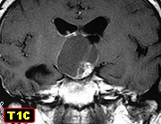
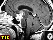
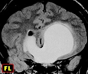
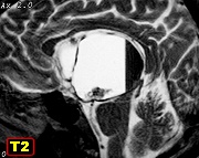
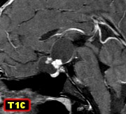
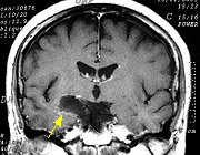
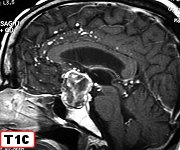
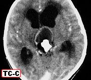
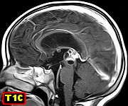
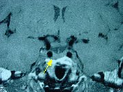
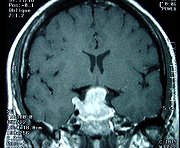
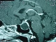
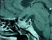
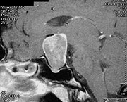
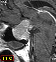
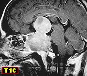
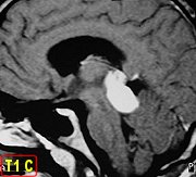
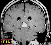
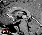
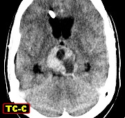
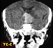
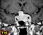
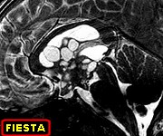
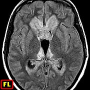
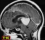
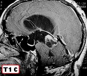
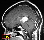
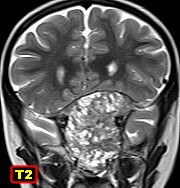
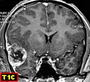
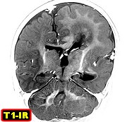
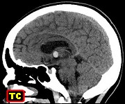
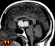
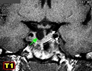
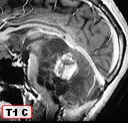
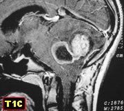
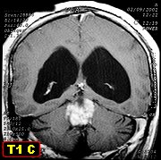
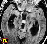
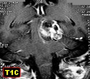
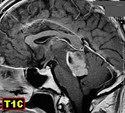
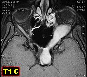
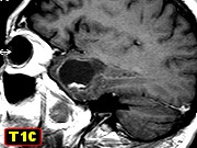
| F. 11 yr.
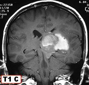
Pilocytic astrocytoma of hypothalamic region |
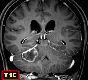
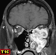
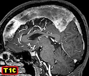
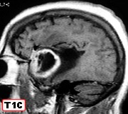
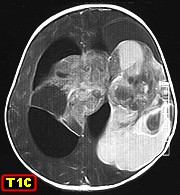
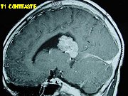
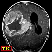
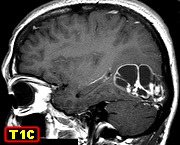
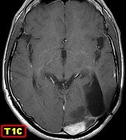
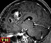
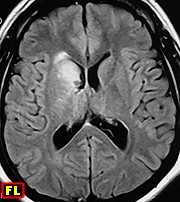
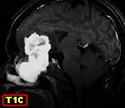
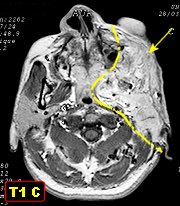
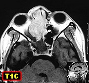
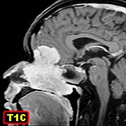
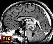
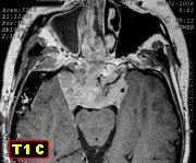
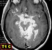
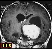
M. 21 yr.
Pilocytic astrocytoma of optic chiasm and hypothalamic region, anaplastic
recurrence |
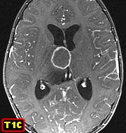
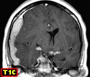
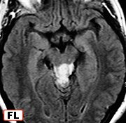
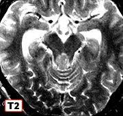
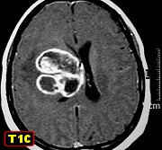
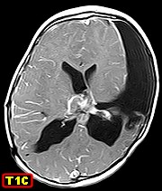
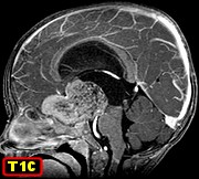
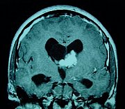
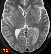
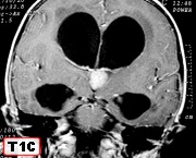
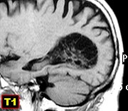
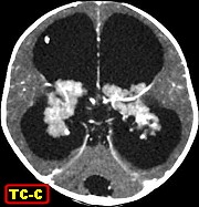
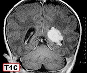
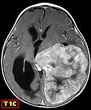
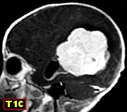
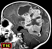
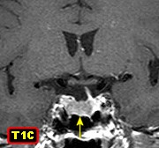
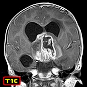
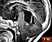
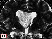
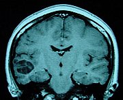
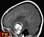
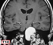
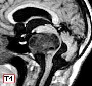
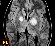
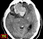
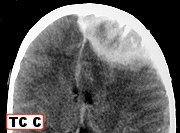
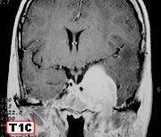
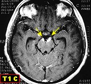
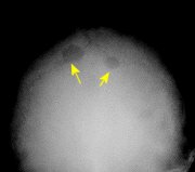
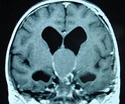
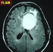
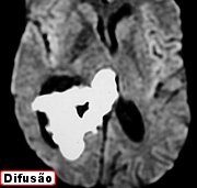
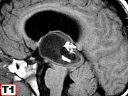
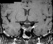
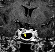
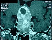
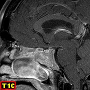
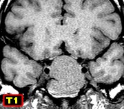
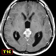
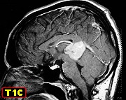
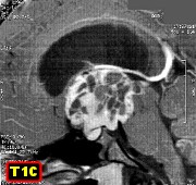
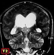
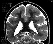
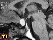
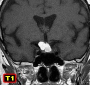
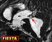
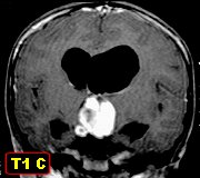
F. 6 yr.
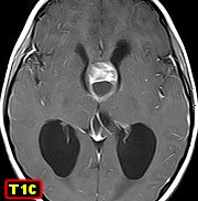
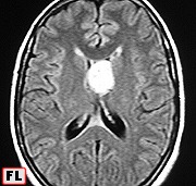
Pilocytic astrocytoma of optic chiasm growing into third ventricle |
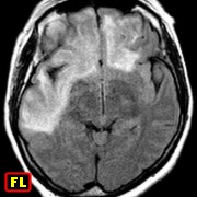
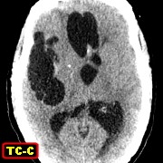
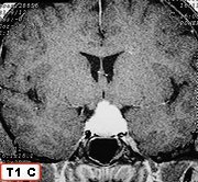
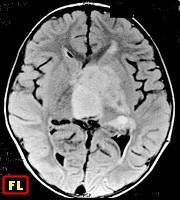
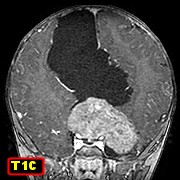
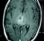
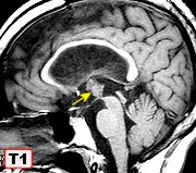
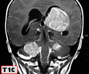
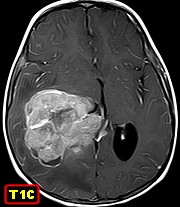
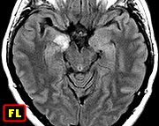
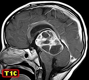
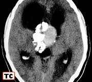
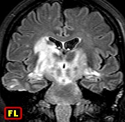
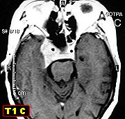
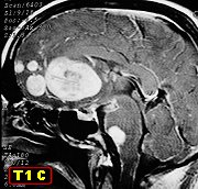
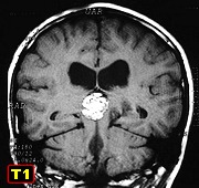
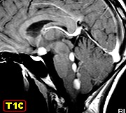
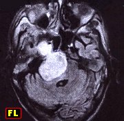
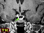
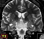
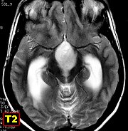
F. 25 yr.
Pilocytic astrocytoma of optic chiasm with third ventricle extension |
|
|
|
|
 |
 |
 |
 |
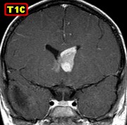
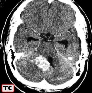
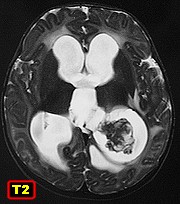
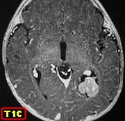
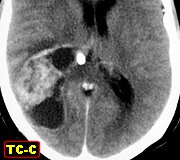
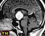
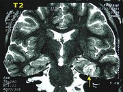
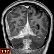
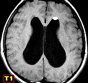
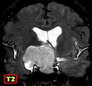
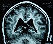
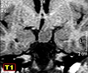
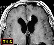
| F. 21 yr.
Pilocytic astrocytoma of chiasmatic region and third ventricle |
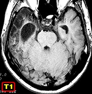
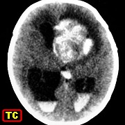
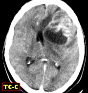
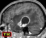
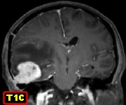
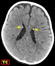
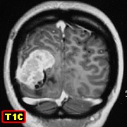
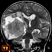
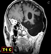
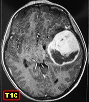
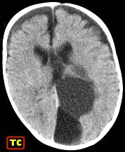
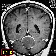
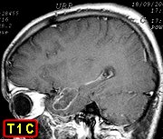
M. 12 yr.
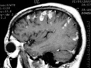
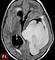
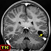
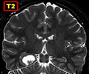
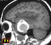
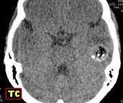
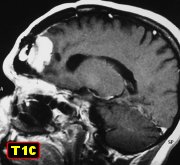
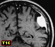
Pilocytic astrocytoma of basal cerebral hemisphere |
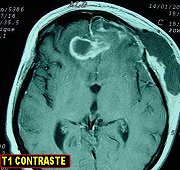
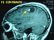
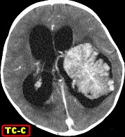
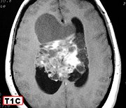
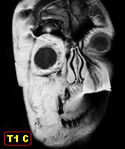
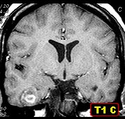
F. 6 yr.
Pilocytic astrocytoma of basal cerebral hemisphere recurring after 4 months |
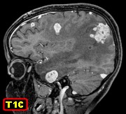
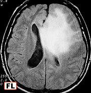
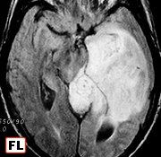
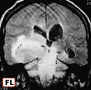
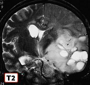
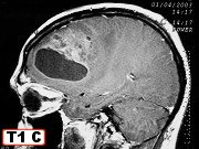
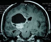
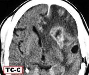
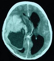
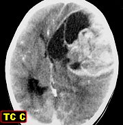
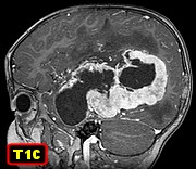
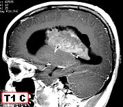
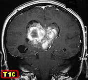
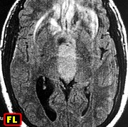
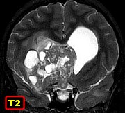
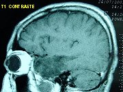
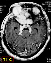
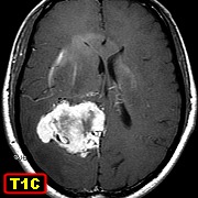
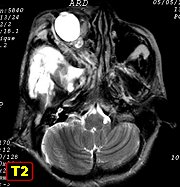
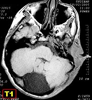
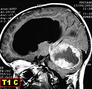
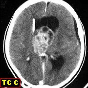
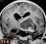
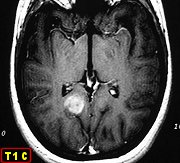
F. 49
yr. Pilocytic astrocytoma of deep cerebral hemisphere with explosive
growth in 4 months |
|
|
|
|
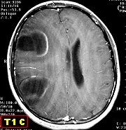
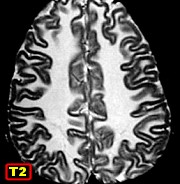
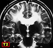
| Cerebral
hemispheres (cortical-subcortical) |
 |
 |
 |
 |
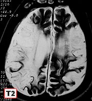
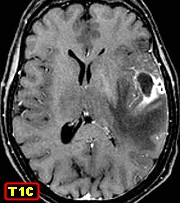
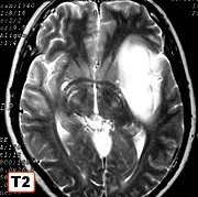
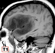
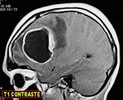
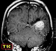
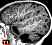
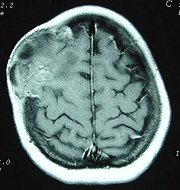
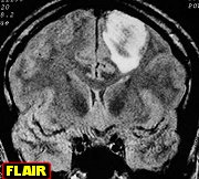
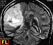
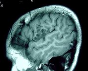
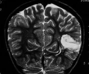
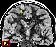
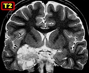
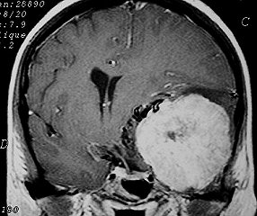
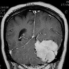
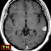
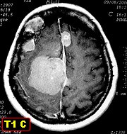
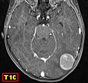
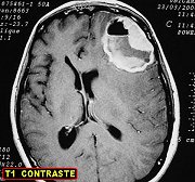
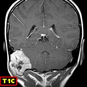
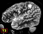
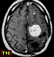
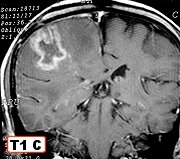
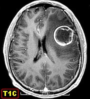
| M. 16 yr.
Right temporal pilocytic astrocytoma |
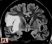
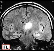
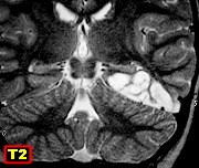
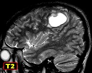
F. 15 yr.
Right temporal pilocytic astrocytoma |
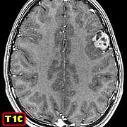
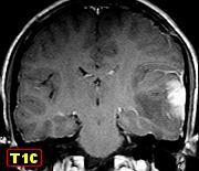
F. 15 yr.
Pilocytic astrocytoma of isthmus of cingulate gyrus |
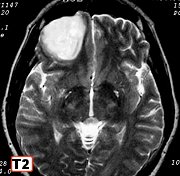
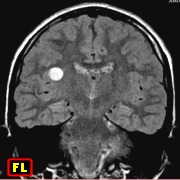
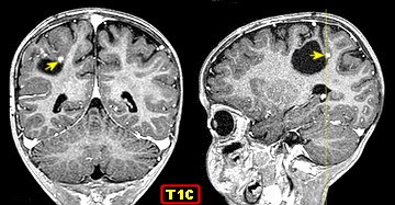
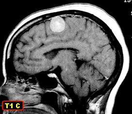
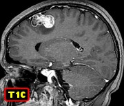
M. 21 yr.
Pilocytic astrocytoma of frontal pole |
|
|
|
|
 |
 |
 |
 |
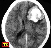
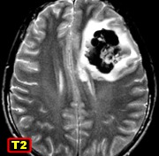
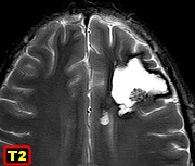
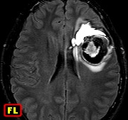
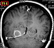
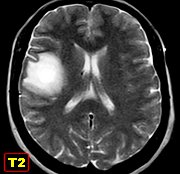
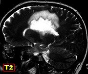
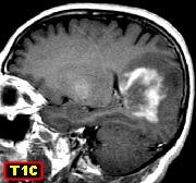
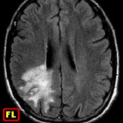
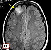
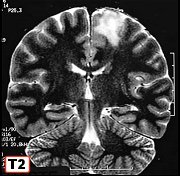
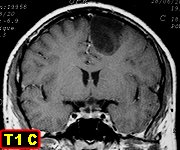
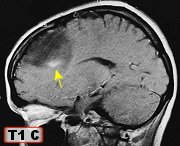
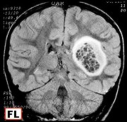
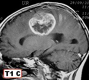
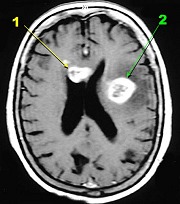
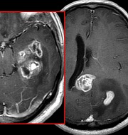
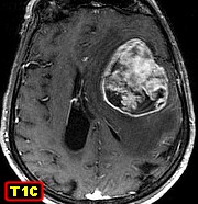
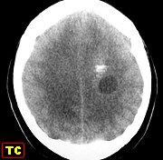
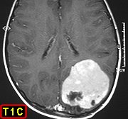
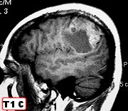
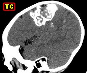
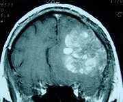
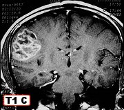
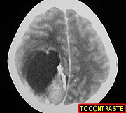
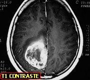
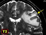
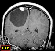
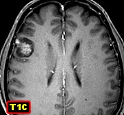
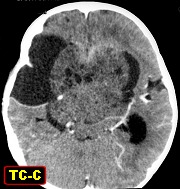
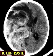
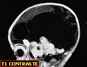
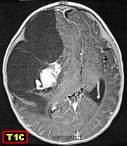
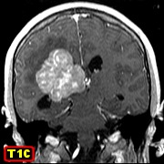
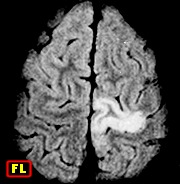
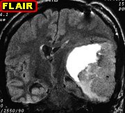
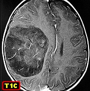
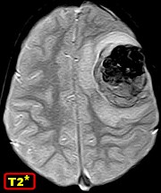
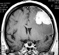
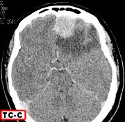
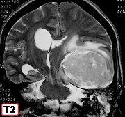
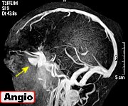
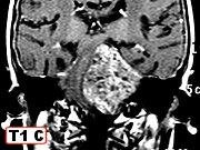
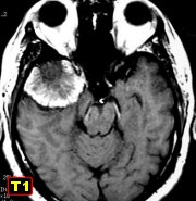
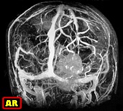
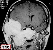
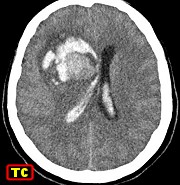
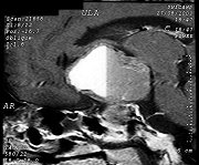
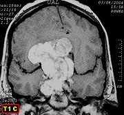
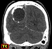
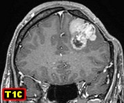
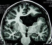
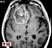
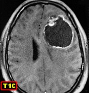
M. 46 yr.
Cystic frontal pilocytic astrocytoma
with abundant
vascular proliferation |
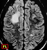
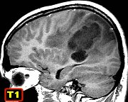
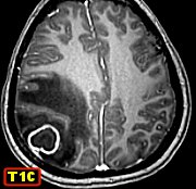
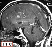
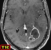
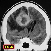
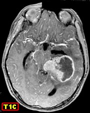
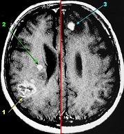
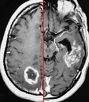
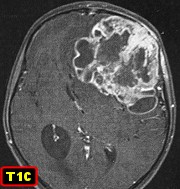
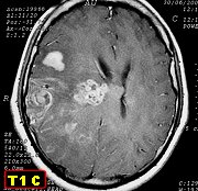
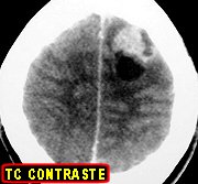
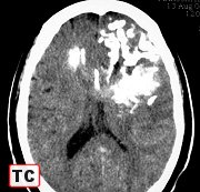
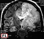
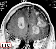
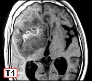
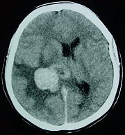
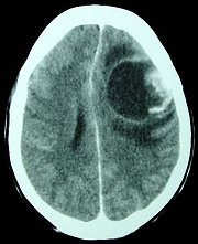
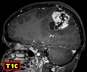
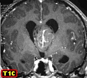
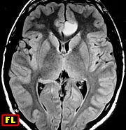
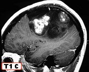
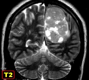
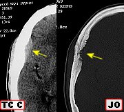
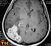
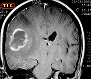
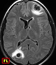
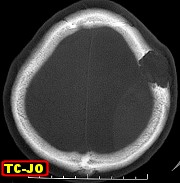
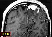
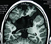
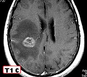
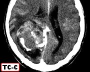
M. 62 yr.
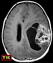
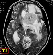
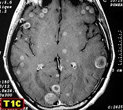
Anaplastic pilocytic astrocytoma of right frontal lobe |
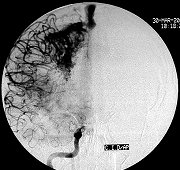
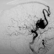
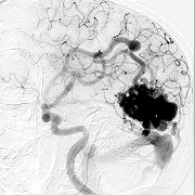
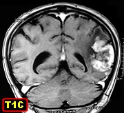
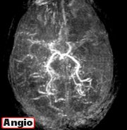
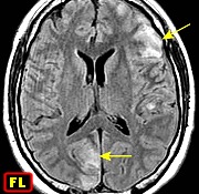
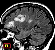
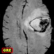
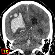
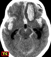
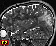
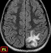
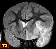
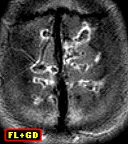
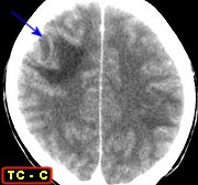
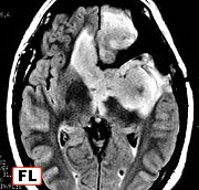
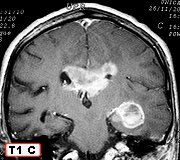
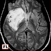
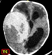
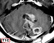
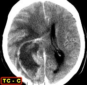
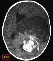
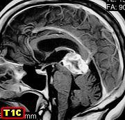
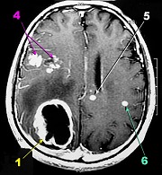
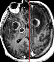
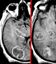
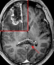
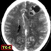
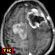
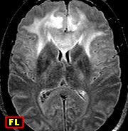
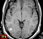
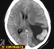
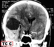
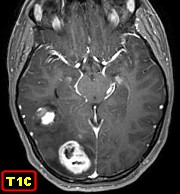
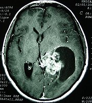
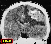
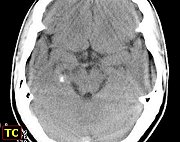
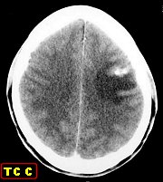
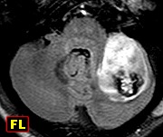
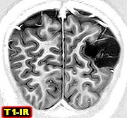
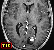
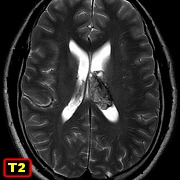
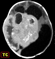
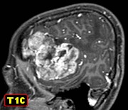
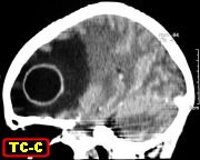
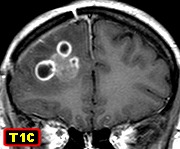
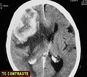
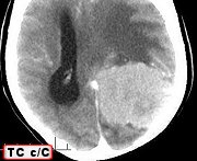
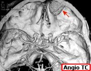
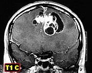
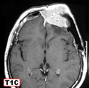
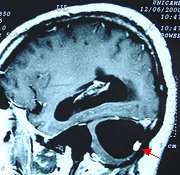
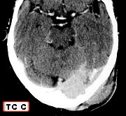
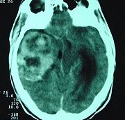
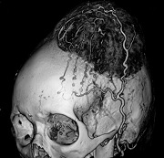
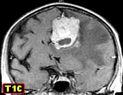
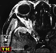
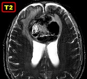
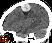
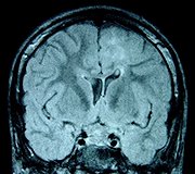
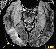
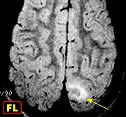
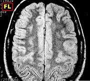
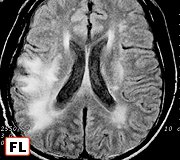
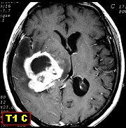
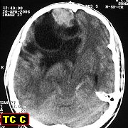
M.
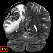
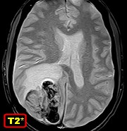
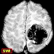
24 yr. Occipital pilocytic astrocytoma featuring grossly abnormal vessels,
leptomeningeal and parenchymal infiltration and ventricular seeding. Case
summary. Text:
pilocytic astrocytomas |
|
|
|
|
|
|
|
Intraventricular |
 |
 |
|
 |
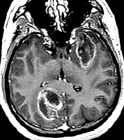
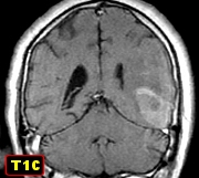
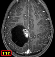
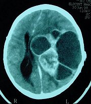
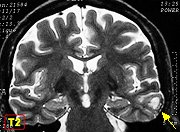
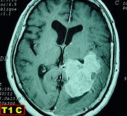
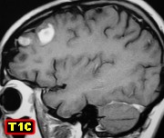
| M. 55 yr.
Temporal pilocytic astrocytoma |
M. 18 yr
9 m. Left frontal pilocytic astrocytoma with pseudo-oligodendroglial
histological pattern |
|
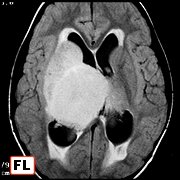
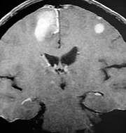
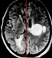
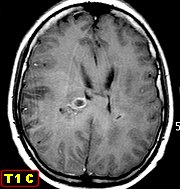
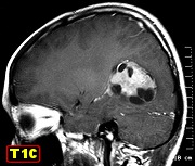
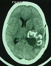
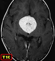
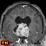
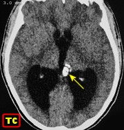
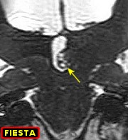
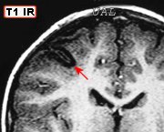
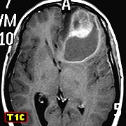
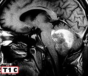
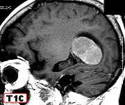
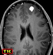
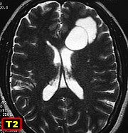
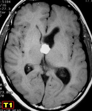
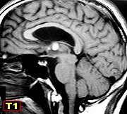
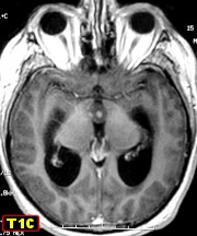
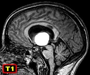
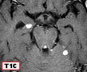
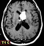
M. 37 yr.
Intraventricular
pilocytic
astrocytoma |
|
|
|
|
| Spinal |
 |
 |
 |
 |
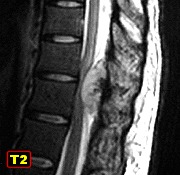
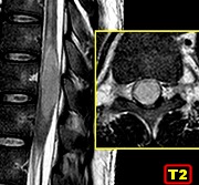
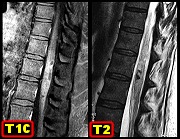
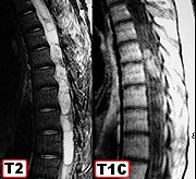
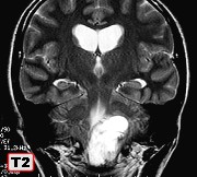
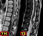
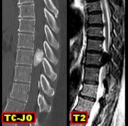
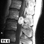
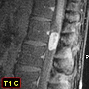
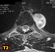
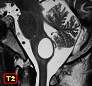
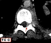
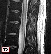
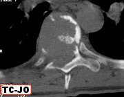
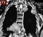
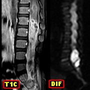
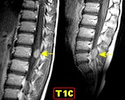
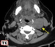
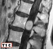
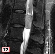
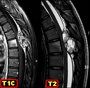
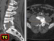
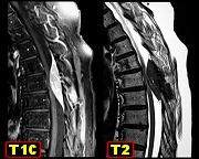
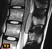
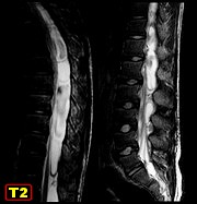
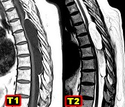
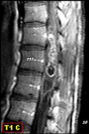
| M. 38 yr.
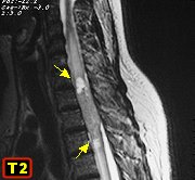
Pilocytic astrocytoma of thoracic/lumbar spinal cord |
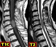
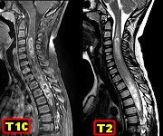
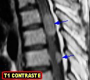
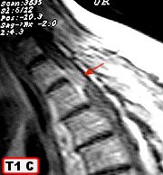
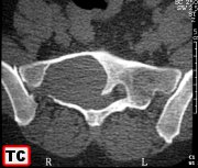
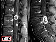
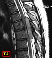
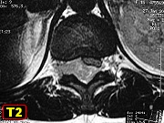
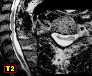
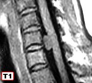
F. 10 yr.
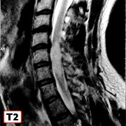
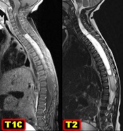
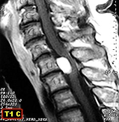
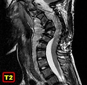
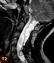
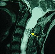
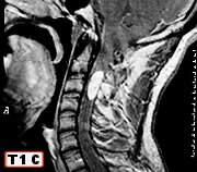
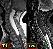
Pilocytic astrocytoma of cervical/thoracic spinal cord |
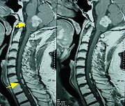
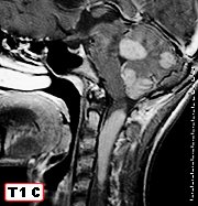
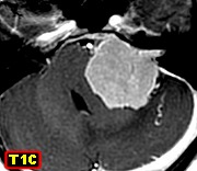
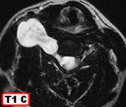
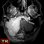
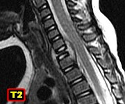
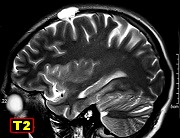
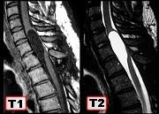
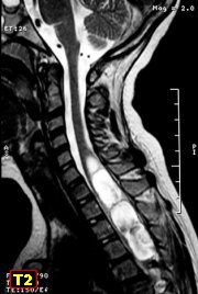
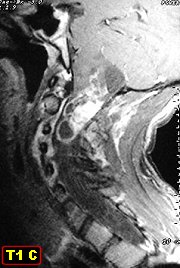
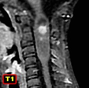
M. 28 yr.
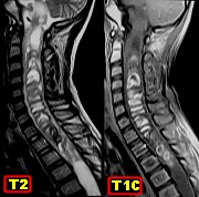
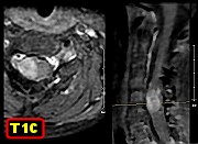
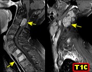
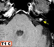
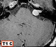
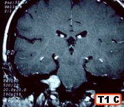
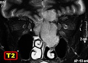
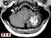
Pilocytic astrocytoma of brain stem and cervical spinal cord |
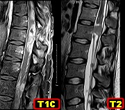
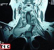
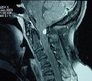
M. 14 yr
3 m. Pilocytic astrocytoma of cervical spinal cord in NF1 |
|
|
|
|