| Normal
nervous tissue and basic pathological reactions |
 |
 |
 |
 |
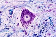
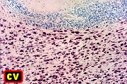
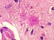
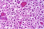
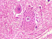
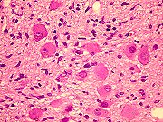
| Motor cerebral
cortex, HE. Pictured - giant pyramidal neurons of Betz |
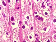
Visual
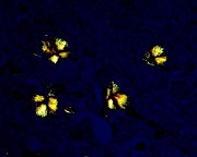
cerebral cortex, HE. Pictured - granular neurons and columns of afferent
axons |
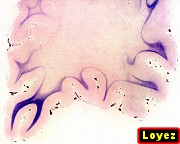
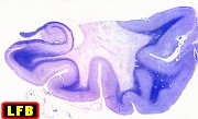
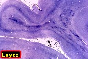
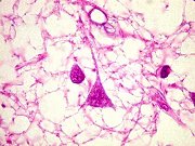
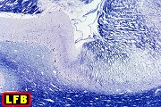
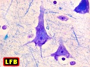
Cerebral
cortex, LFB - Nissl |
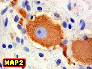
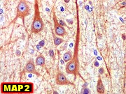
Cerebral
cortex, IH for MAP2 |
|
|
|
|
 |
 |
 |
 |
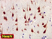
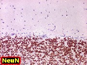
| Cerebral
cortex, IH for NeuN |
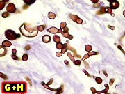
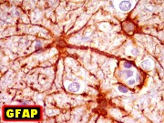
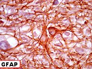
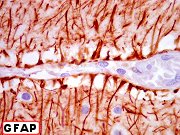
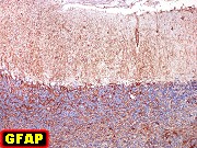
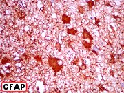
Cerebral
cortex, IH for GFAP |
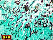
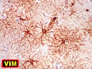
Cerebral
cortex, IH for VIM |
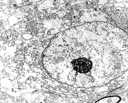
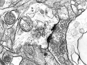
Cerebral
cortex, electron microscopy |
|
|
|
|
 |
 |
 |
 |
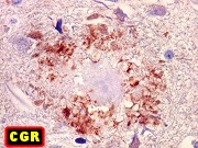
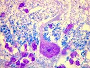
| Medulla
oblongata - immunohistochemistry |
Pineal
- HE, IH |
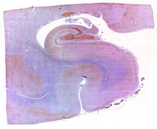
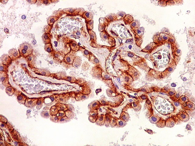
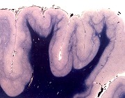
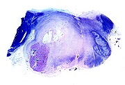
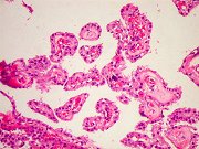
Choroid
plexus and ependyma from autopsy. HE, IH |
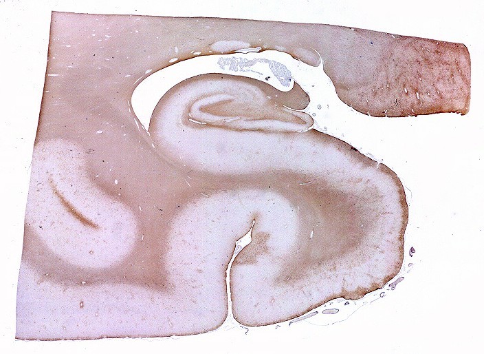
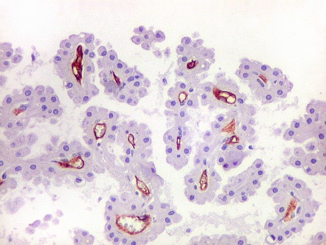
Choroid
plexus - biopsy. HE, IH |
|
|
|
|
 |
 |
 |
 |
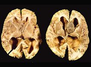
| Cerebellum
- HE |
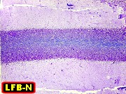
Cerebellum
(dog) - LFB/Nissl |
Cerebellum
- IH for glial antigens: GFAP, VIM, S100 |
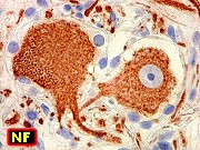
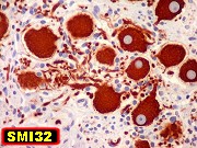
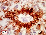
Cerebellum
- IH for neuronal antigens: NSE, NF, SNF |
|
|
|
|
 |
 |
 |
 |
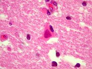
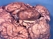
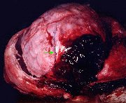
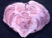
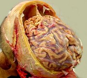
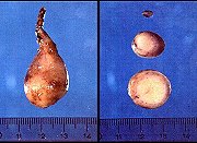
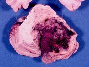
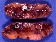
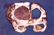
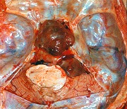
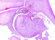
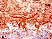
| M. 22 yr.
Lhermitte-Duclos
disease, area of normal cerebellum. HE, LFB-Nissl |
Same, IH
for MAP2, NeuN, NF, synaptophysin |
Same, IH
for GFAP, S100, CD34 |
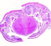
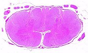
Spinal
cord |
|
|
|
|
 |
 |
 |
 |
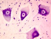
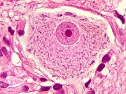
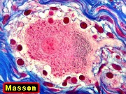
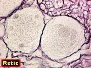
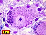
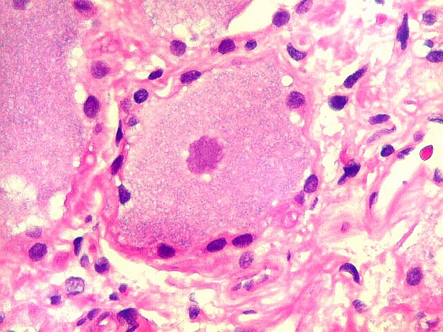
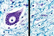
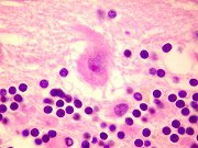
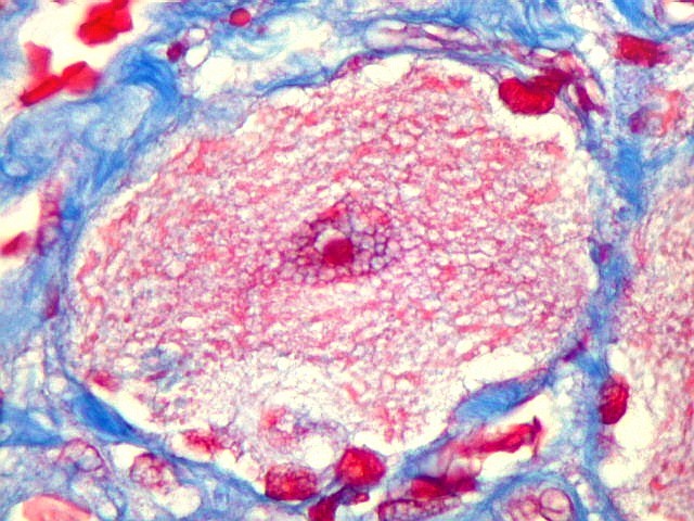
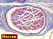
| Normal
sensory ganglion, HE, Masson, reticulin |
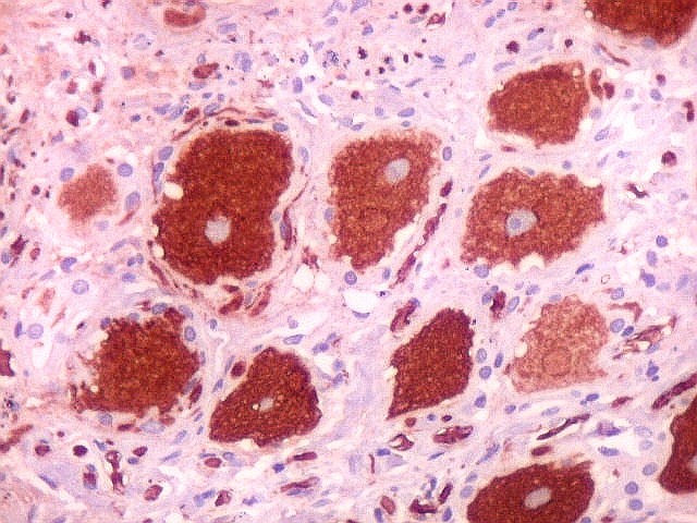
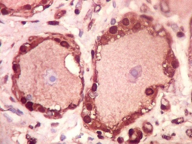
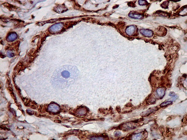
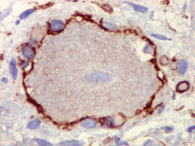
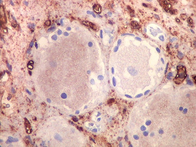
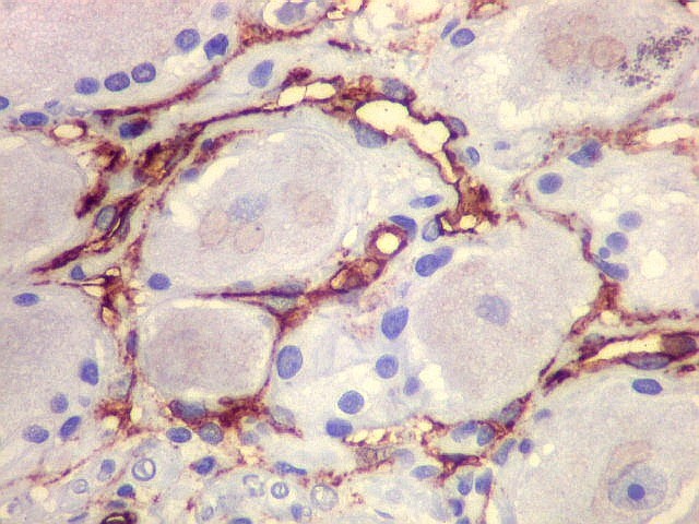
Normal
sensory ganglion, IH |
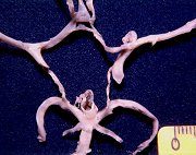
Cauda equina |
Spinal
nerve root |
|
|
|
|
 |
 |
 |
 |
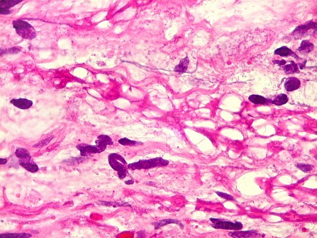
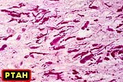
| Filum terminale |
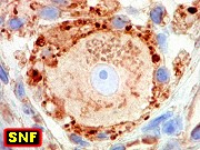
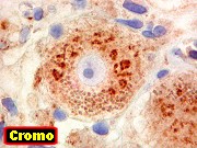
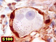
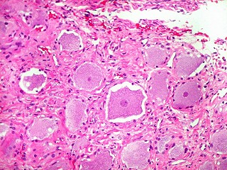
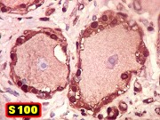
Sympathetic
ganglion |
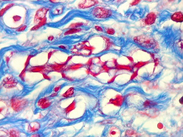
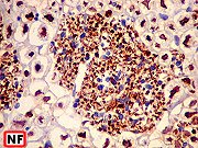
Normal
human median nerve. HE, Masson's trichrome, IH for neurofilament
and S100 |
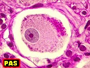
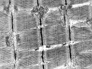
Normal
small nerve of ocular conjunctiva, electron microscopy |
|
|
|
|
 |
 |
 |
 |
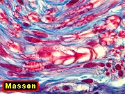
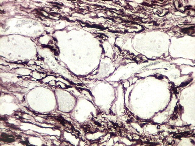
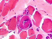
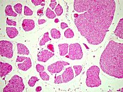
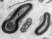
| Normal
human skeletal muscle, electron microscopy |
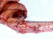
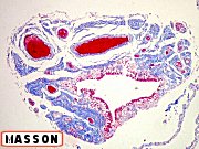
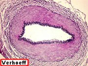
Normal
temporal artery, HE, Masson's trichrome, Verhoeff |
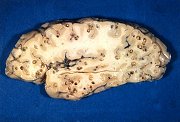
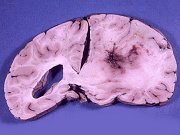
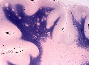
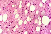
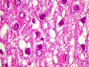
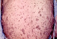
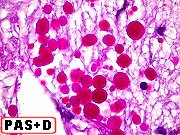
Corpora
amylacea |
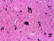
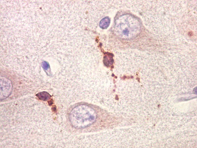
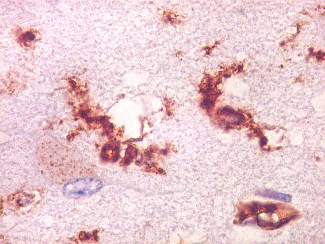
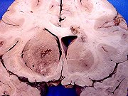
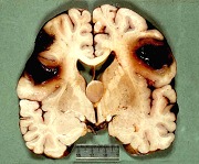
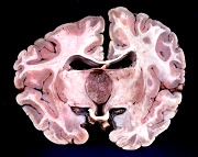
Periventricular
leukomalacia, HE, GFAP, VIM |
|
|
|
|