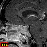
| 17/2/12 |
| New
Cases of Neuroimaging & Neuropathology: |
 |
 |
 |
 |
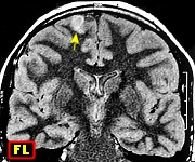
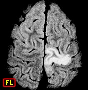
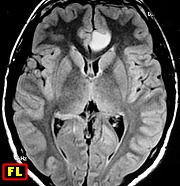
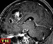
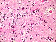
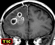
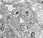
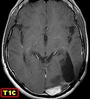
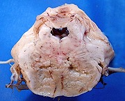
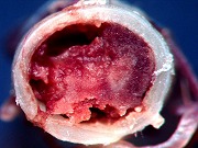
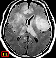
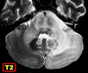
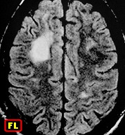
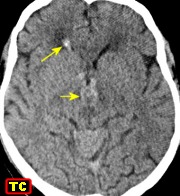
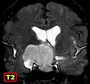
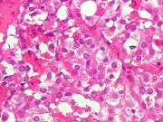
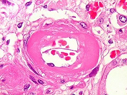
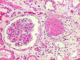
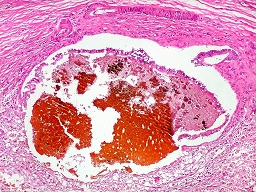
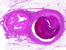
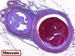
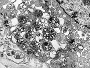
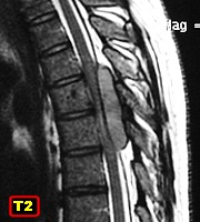
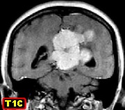
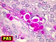
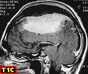
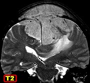
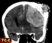
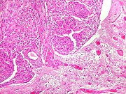
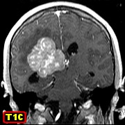
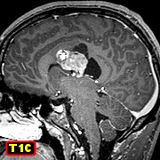
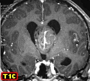
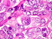
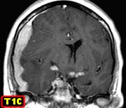
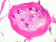
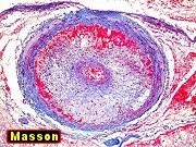
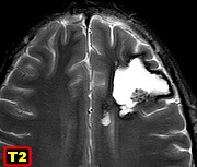
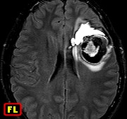
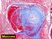
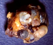
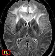
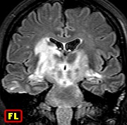
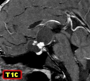
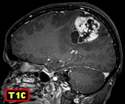
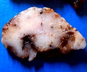
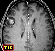
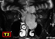
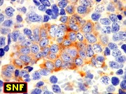
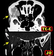
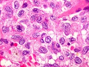
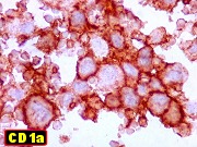
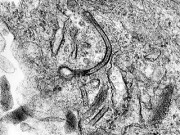
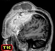
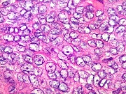
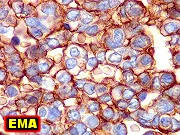
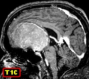
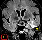
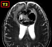
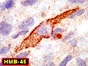
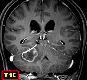
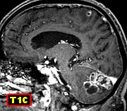
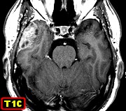
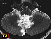
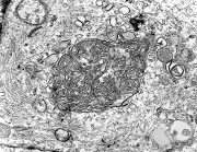
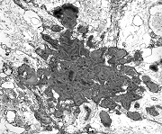
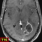
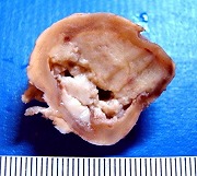
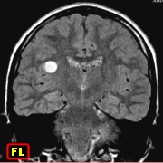
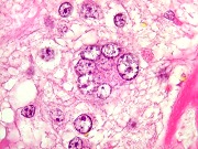
| F. 58 yr.
Giant cell glioblastoma with a component of conventional glioblastoma multiforme |
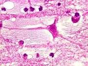
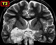
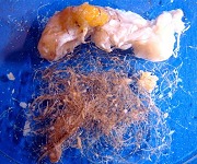
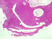
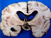
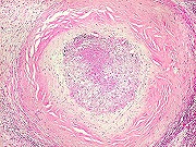
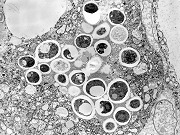
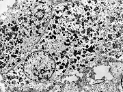
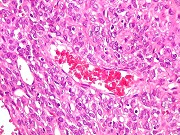
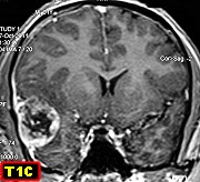
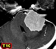
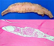
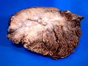
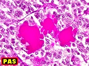
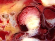
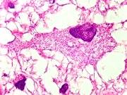
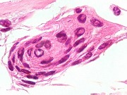
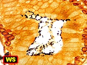
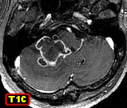
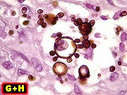
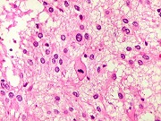
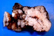
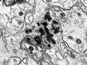
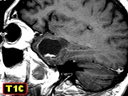
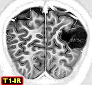
Same, surgical
specimen. Encapsulated tumor with necrotic center |
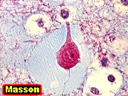
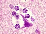
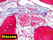
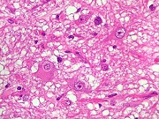
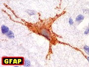
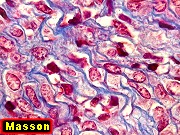
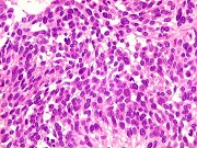
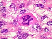
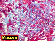
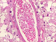
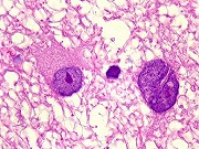
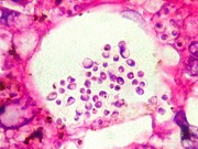
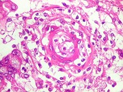
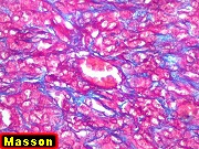
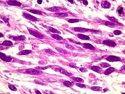
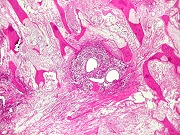
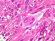
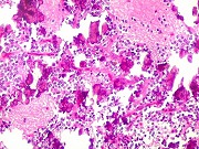
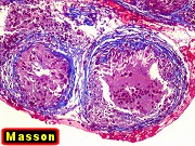
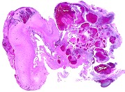
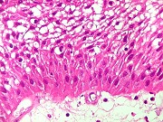
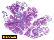
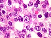
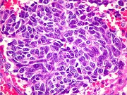
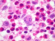
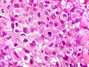
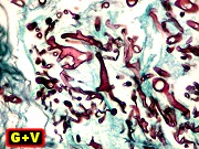
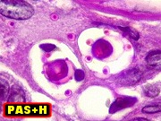
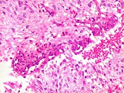
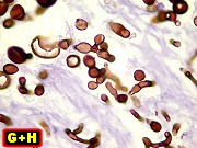
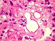
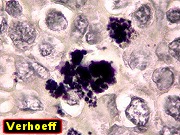
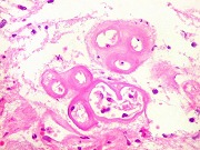
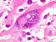
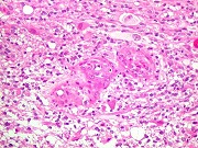
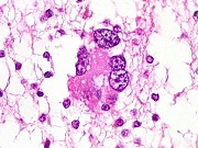
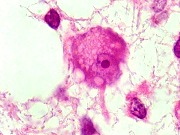
Same, giant
cell part - HE, Masson's trichrome |
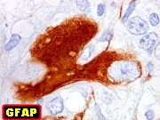
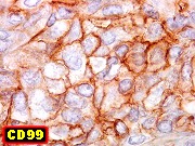
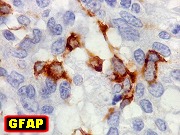
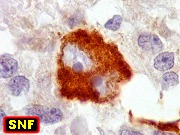
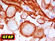
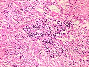
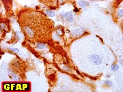
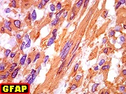
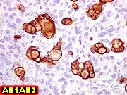
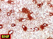
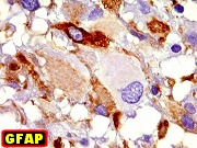
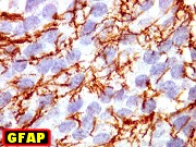
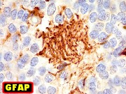
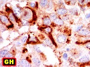
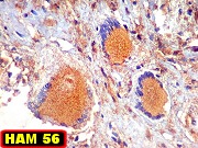
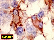
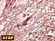
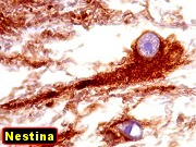
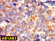
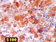
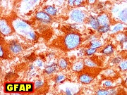
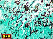
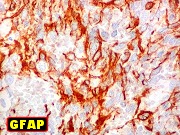
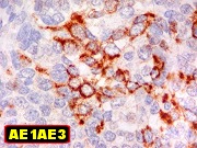
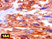
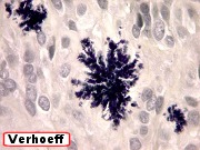
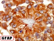
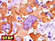
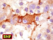
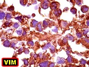
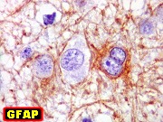
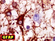
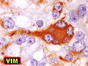
Same, giant
cell part - IH - glial markers, GFAP, VIM, S-100 |
|
|
|
|
 |
 |
 |
 |
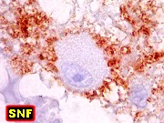
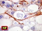
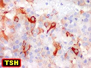
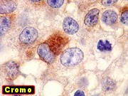
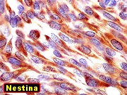
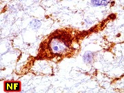
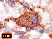
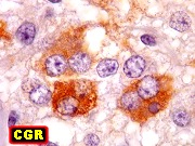
| Same, giant
cell part - IH - neuronal markers, synaptophysin, chromogranin, NF, class
III beta-tubulin |
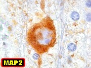
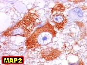
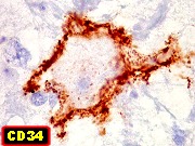
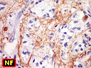
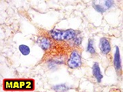
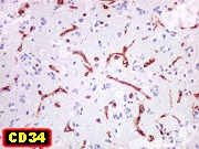
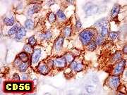
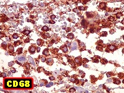
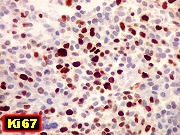
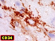
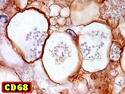
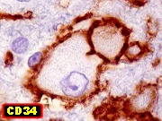
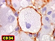
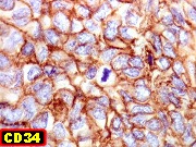
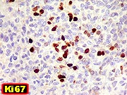
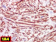
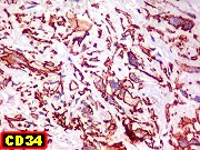
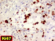
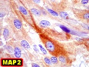
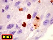
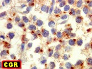
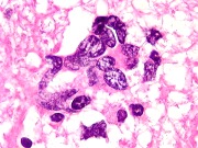
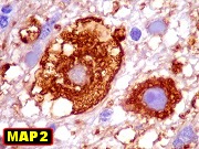
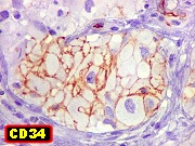
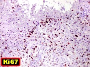
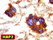
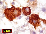
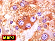
Same, giant
cell part - IH - MAP2, CD34, Ki-67, p53 |
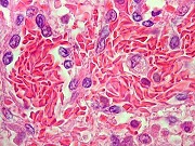
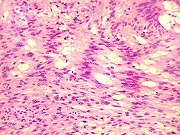
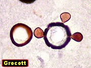
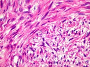
Same, conventional
glioblastoma part - HE |
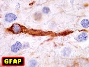
Same, conventional
glioblastoma part - IH - glial markers, GFAP, VIM |
|
|
|
|
 |
 |
|
 |
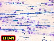
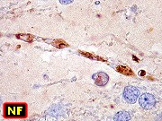
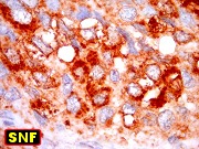
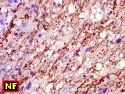
| Same, conventional
glioblastoma part - IH - neuronal markers, NF, MAP2, tubulin. Pictured
- neoplastic infiltration among axons |
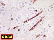
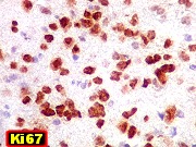
Same, conventional
glioblastoma part - IH - CD34, Ki-67, p53 |
|
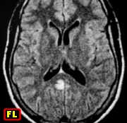
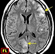
F. 45 yr.
Left parietal ganglioglioma |
|
|
|
|
 |
 |
 |
 |
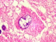
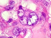
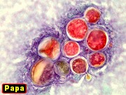
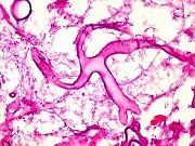
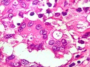
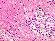
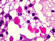
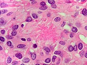
| Same, HE.
Pictured - multinucleated tumor cell |
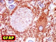
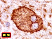
Same, IH
- glial markers, GFAP, VIM |
Same, IH
- neuronal markers, synaptophysin, NF, chromogranin, NeuN |
Same, IH
- MAP2, tubulin, CD34, Ki-67 |
|
|
|
|
 |
 |
 |
 |
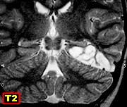
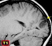
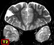
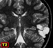
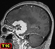
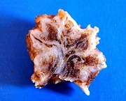
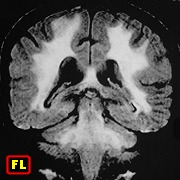
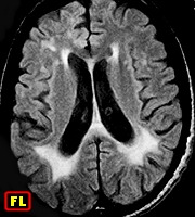
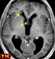
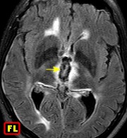
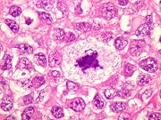
| M. 12 yr.
Small solid ganglioglioma of right insula |
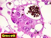
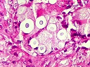
Same, HE.
Pictured - abnormal neuron |
Same, IH
- glial markers, GFAP, VIM |
Same, IH
- neuronal markers, synaptophysin, NF, chromogranin, NeuN |
|
|
|
|
 |
|
 |
 |
| Same, IH
- MAP2, tubulin, CD34, Ki-67 |
|
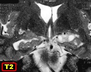
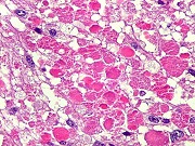
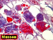
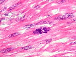
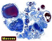
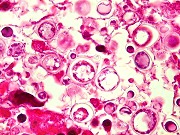
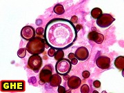
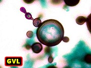
F. 51 yr.
Solid-cystic ganglioglioma of right temporal and occipital regions |
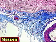
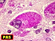
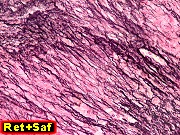
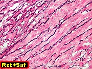
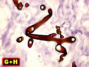
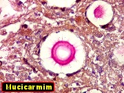
Same, HE,
Masson's trichrome. Pictured - multinucleated tumor cell |
|
|
|
|
 |
 |
 |
|
| Same, IH
- glial markers, GFAP, VIM |
Same, IH
- neuronal markers, synaptophysin, chromogranin, NF, NeuN |
Same, IH
- MAP2, tubulin, CD34, Ki-67 |
|