| 4/11/15 |
| New
Cases of Neuroimaging & Neuropathology: |
 |
 |
 |
 |
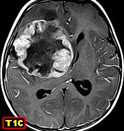
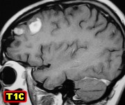
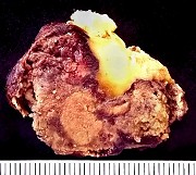
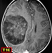
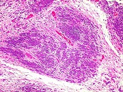
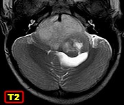
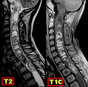
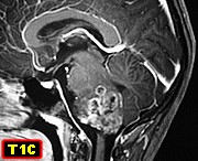
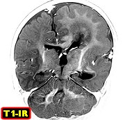
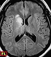
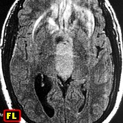
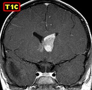
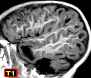
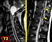
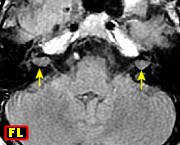
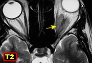
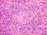
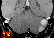
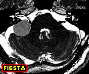
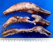
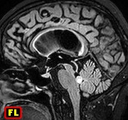
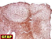
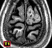
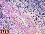
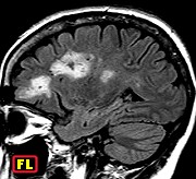
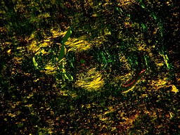
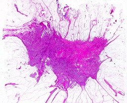
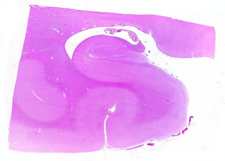
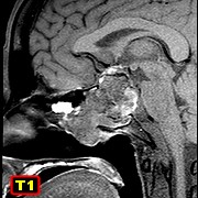
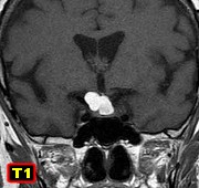
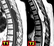
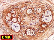
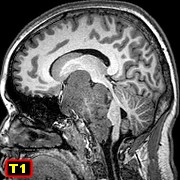
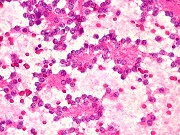
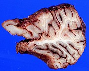
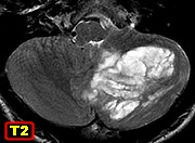
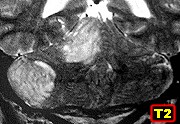
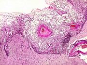
| F. 32 yr.
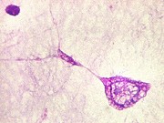
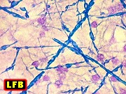
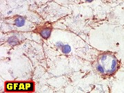
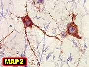
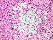
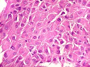
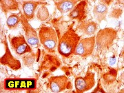
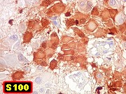
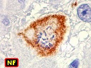
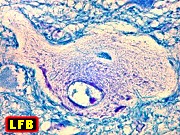
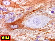
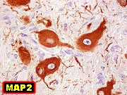
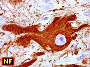
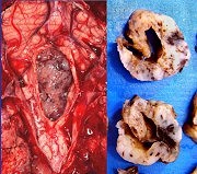
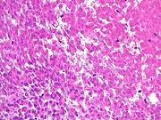
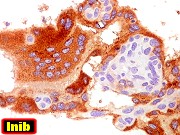
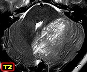
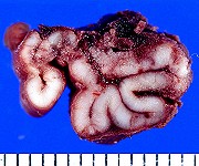
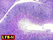
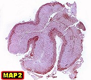
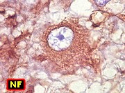
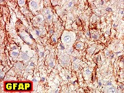
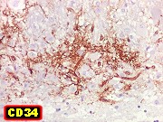
Dysplastic gangliocytoma of cerebellum (Lhermitte-Duclos disease).
MRI |
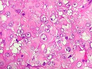
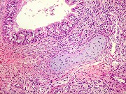
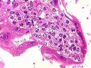
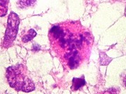
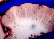
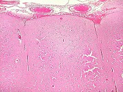
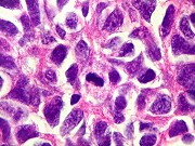
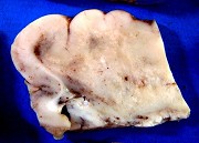
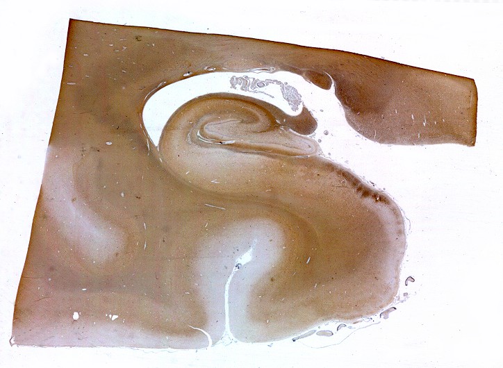
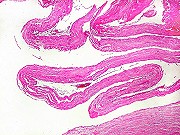
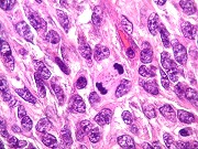
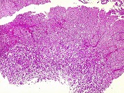
Same, full
blown changes, specimen, HE |
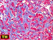
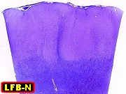
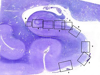
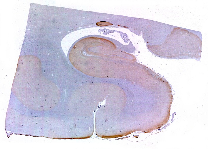
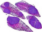
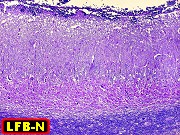
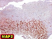
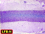
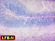
Same,
LFB-Nissl |
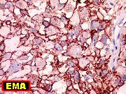
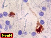
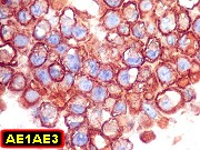
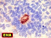
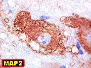
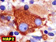
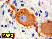
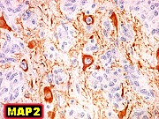
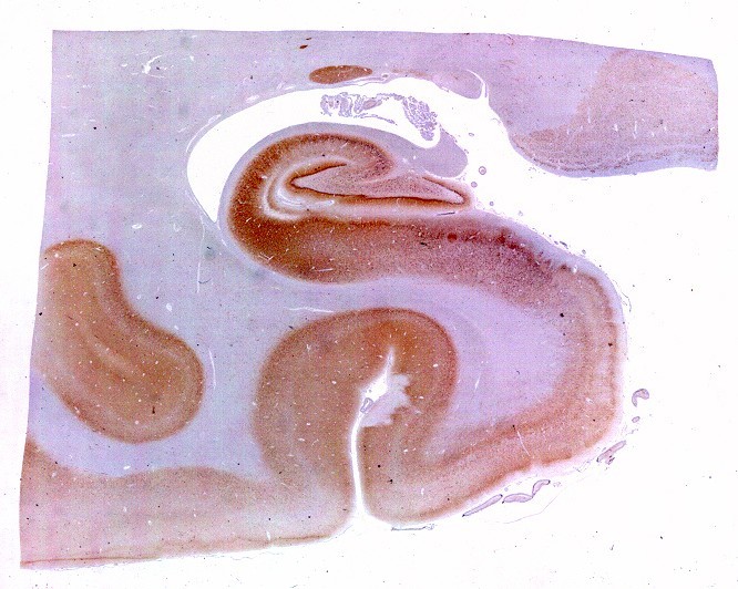
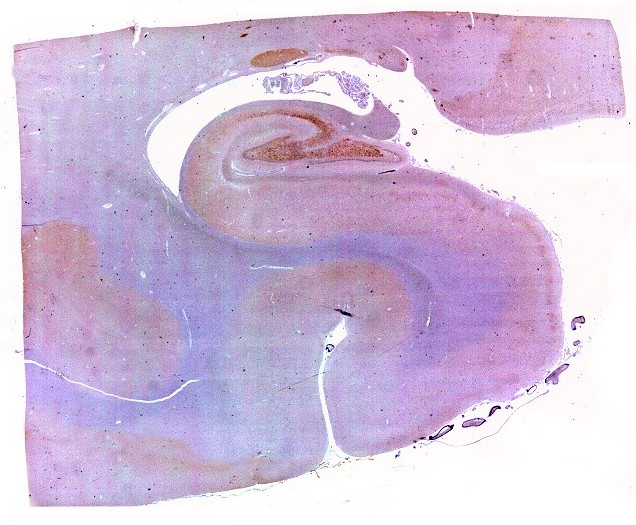
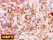
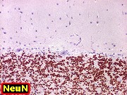
Same,
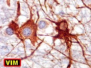
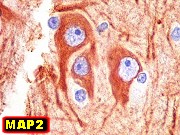
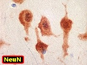
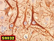
immunohistochemistry for MAP2, NeuN, synaptophysin |
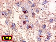
| Lhermitte-Duclos:
image
bank, text |
|
|
 |
 |
 |
 |
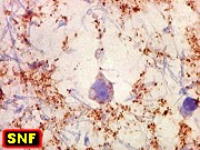
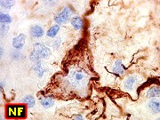
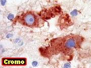
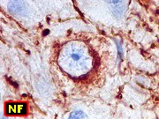
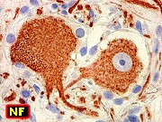
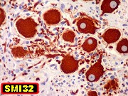
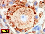
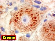
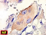
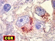
| Same,
IH for NF, chromogranin |
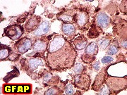
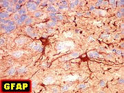
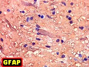
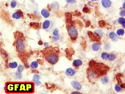
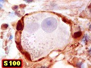
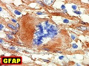
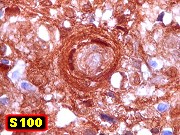
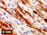
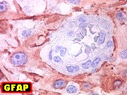
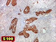
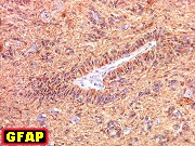
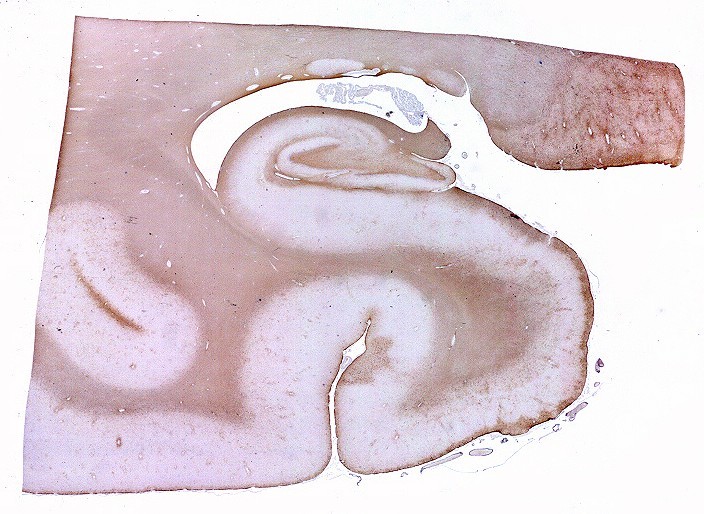
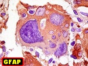
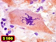
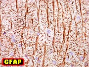
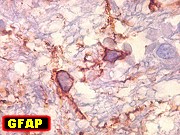
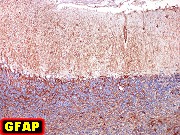
Same, IH
for GFAP, S100 |
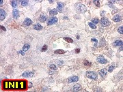
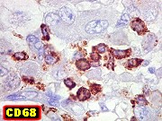
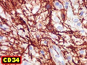
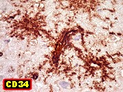
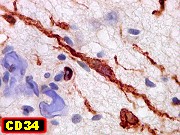
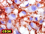
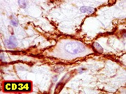
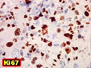
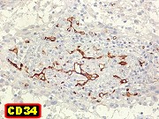
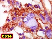
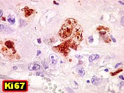
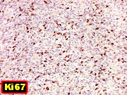
Same,
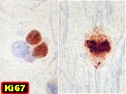
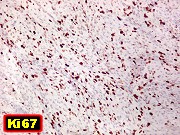
IH for CD34, CD68, Ki67 |
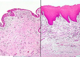
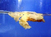
Same, partial
changes, specimen, HE |
|
|
|
|
 |
 |
 |
|
| Same, LFB-Nissl |
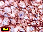
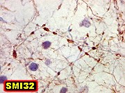
Same, IH
for MAP2, NeuN |
Same, IH
for GFAP, CD34, CD68, Ki67 |
|
|
|
|
|
 |
 |
 |
 |
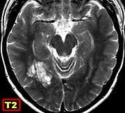
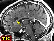
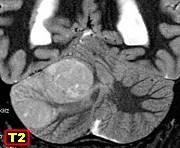
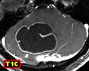
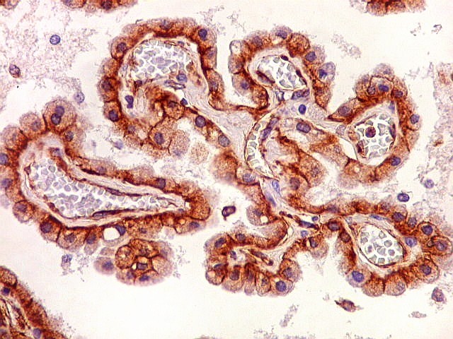
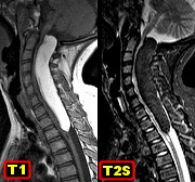
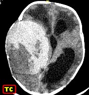
| M. 22 yr.
Dysplastic gangliocytoma of cerebellum (Lhermitte-Duclos disease).
MRI |
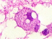
Same, HE,
LFB-Nissl |
Same, IH
for MAP2, NeuN, synaptophysin |
Same, IH
for NF, chromogranin |
|
|
|
|
 |
 |
 |
 |
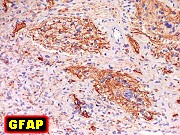
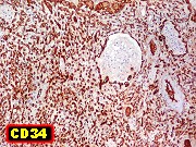
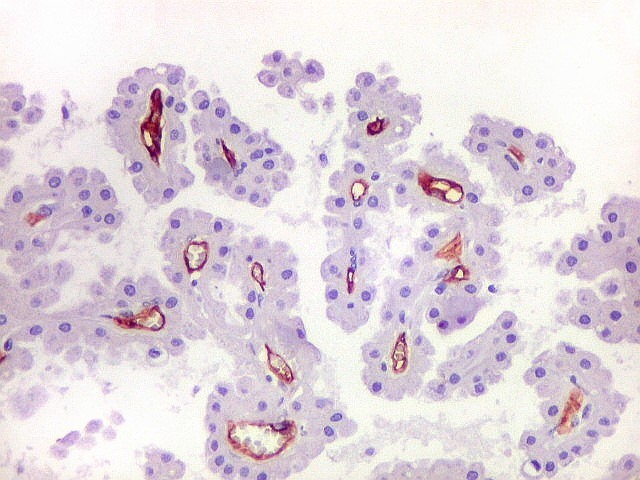
| Same, IH
for GFAP, CD34, Ki67 |
M. 22 yr.
Same patient, normal cerebellar cortex. HE, LFB-Nissl |
Same, IH
for MAP2, NeuN, NF, synaptophysin |
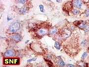
Same, IH
for GFAP, S100, CD34 |
|
|
|
|
 |
 |
|
|
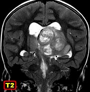
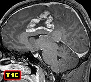
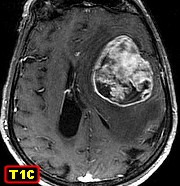
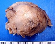
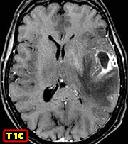
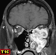
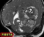
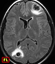
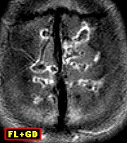
| F. 45 yr.
Dysplastic gangliocytoma of cerebellum (Lhermitte-Duclos disease).
MRI - two distinct lesions in right cerebellar hemisphere |
Same, HE,
LFB-Nissl, lesions at different phases |
|
|
|
|
|
|
 |
 |
|
 |
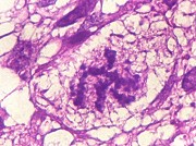
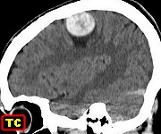
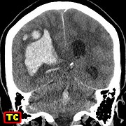
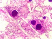
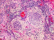
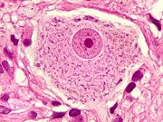
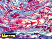
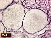
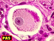
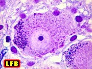
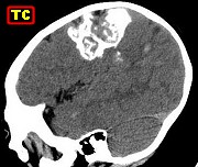
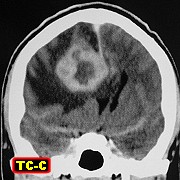
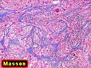
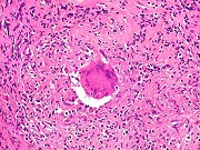
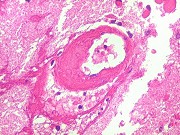
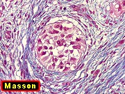
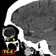
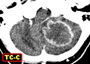
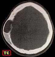
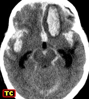
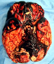
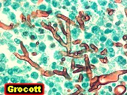
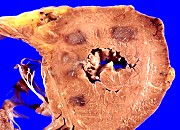
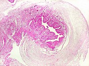
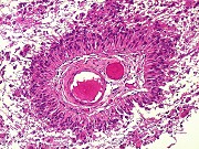
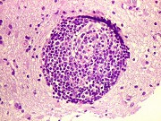
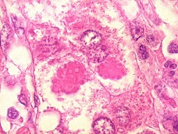
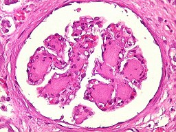
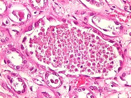
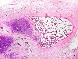
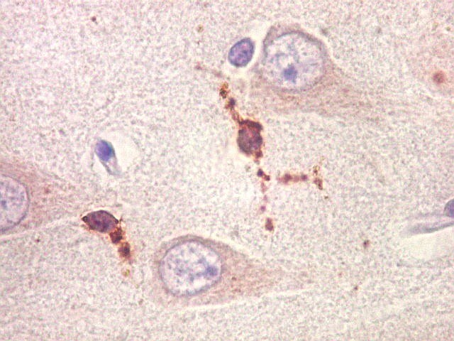
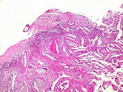
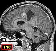
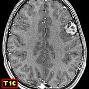
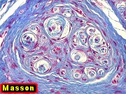
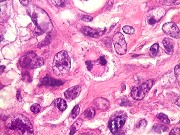
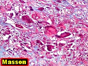
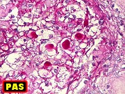
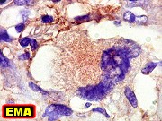
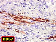
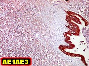
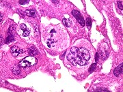
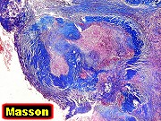
M. 32 yr.
Cerebral granulomatous cryptococcosis:
circumscribed
meningeal, cerebral lesions |
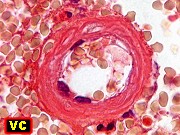
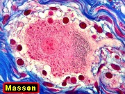
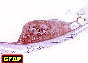
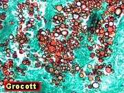
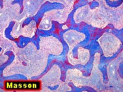
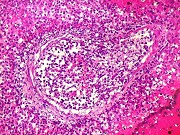
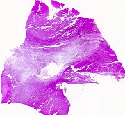
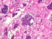
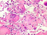
Same, HE,
Masson's trichrome, Grocott, mucicarmine, PAS |
|
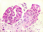
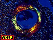
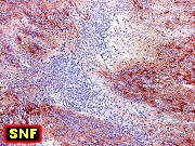
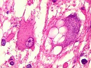
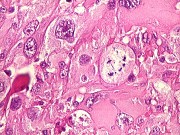
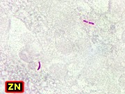
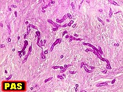
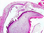
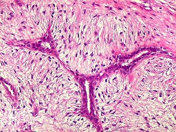
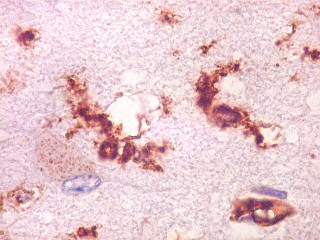
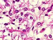
Cerebral
cryptococcosis: non specific and granulomatous inflammation in meninges
and parenchyma. HE, PAS |
|
|
|
|