| 22/8/14 |
| New
Cases of Neuroimaging & Neuropathology: |
 |
 |
 |
|
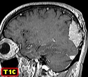
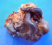
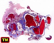
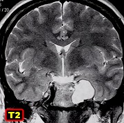
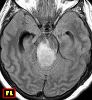
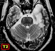
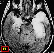
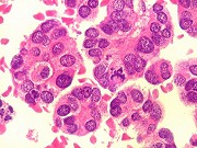
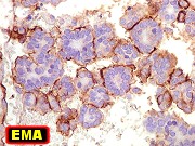
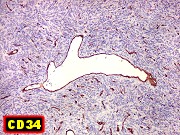
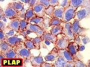
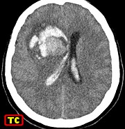
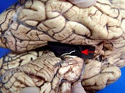
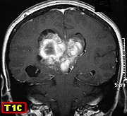
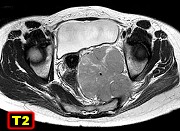
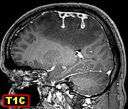
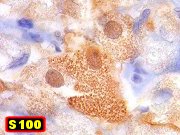
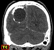
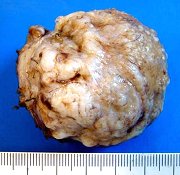
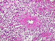
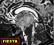
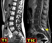
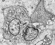
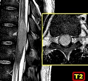
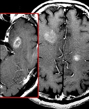
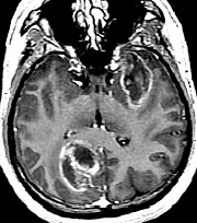
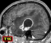
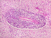
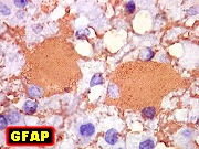
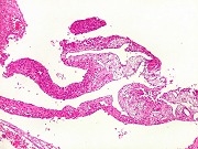
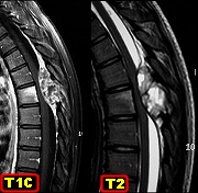
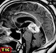
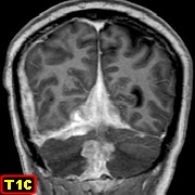
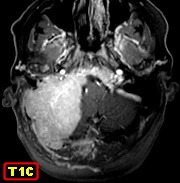
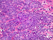
| F. 36 yr.
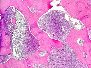
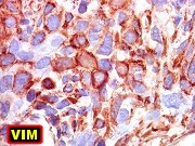
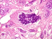
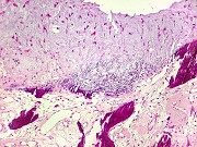
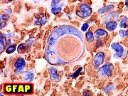
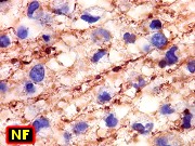
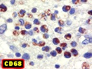
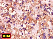
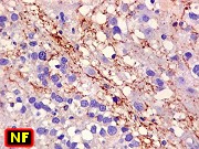
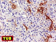
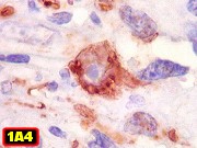
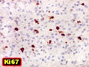
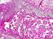
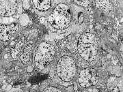
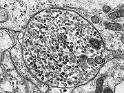
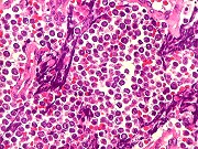
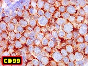
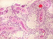
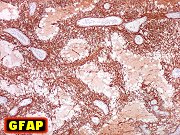
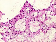
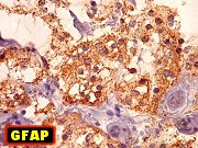
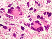
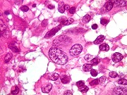
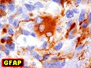
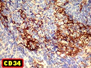
Primitive neuroectodermal tumor (PNET) of brainstem at IV ventricle floor |
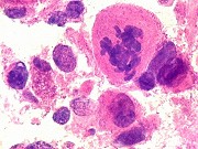
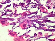
Same, HE |
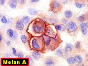
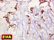
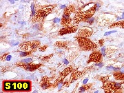
Same, immunohistochemistry |
|
|
|
|
|
 |
 |
 |
|
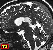
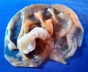
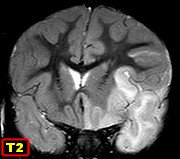
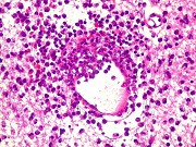
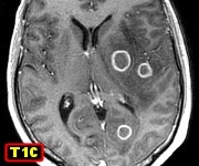
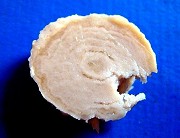
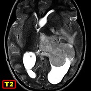
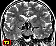
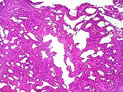
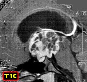
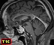
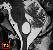
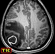
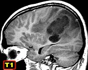
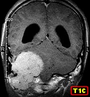
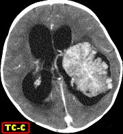
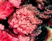
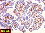
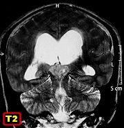
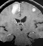
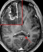
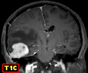
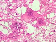
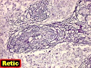
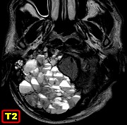
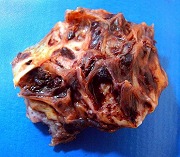
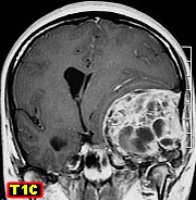
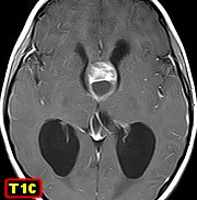
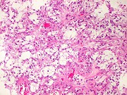
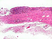
| M. 8 yr.
Pilomyxoid astrocytoma at septum pellucidum |
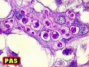
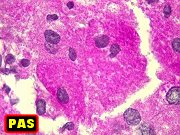
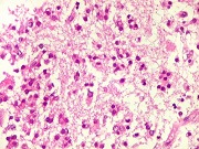
Same, HE,
solid component |
Same, HE,
cyst wall |
|
|
|
|
|
 |
 |
 |
Leukodystrophies
-
Illustrated
linked text (in Portuguese) |
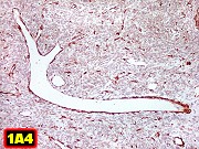
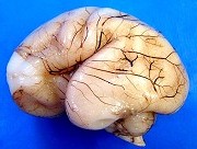
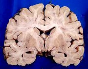
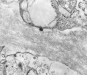
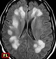
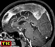
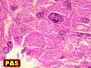
| M. 43 yr.
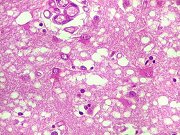
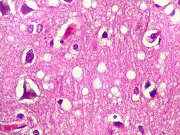
Leukodystrophy with spongiform changes of white matter reminiscent of Canavan's
disease |
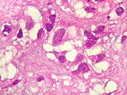
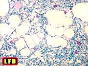
Same, HE,
LFB-Nissl |
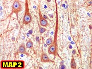
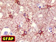
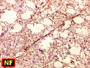
Same, IH |
|
|
|
|
|
 |
 |
 |
 |
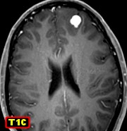
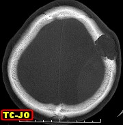
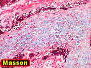
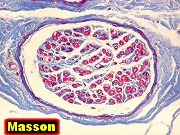
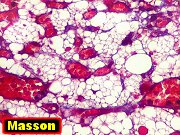
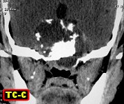
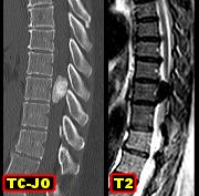
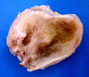
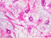
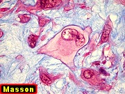
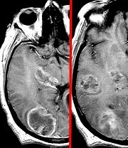
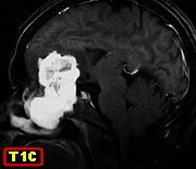
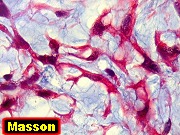
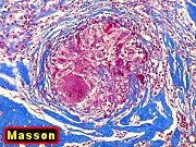
| F. 51 yr.
Sarcoidosis of falx and dura mater of cerebral convexity |
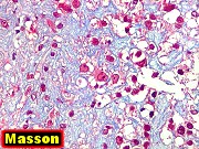
Same, HE,
Masson's trichrome |
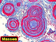
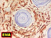
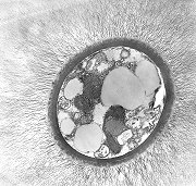
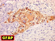
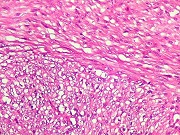
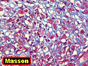
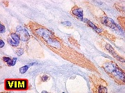
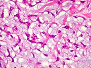
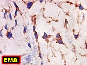
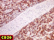
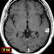
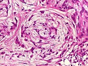
M. 38 yr.
Small temporal chordoid meningioma |
Same, HE,
IH. Pictured - incipient whorl formation |
|
|
|
|
 |
 |
|
|
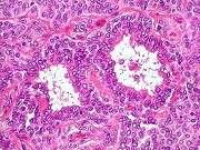
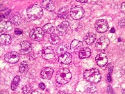
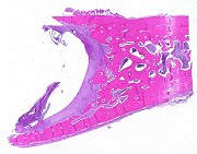
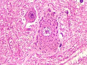
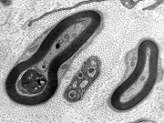
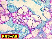
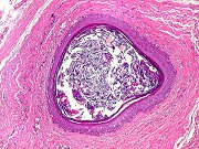
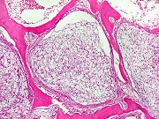
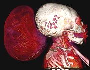
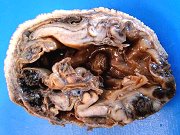
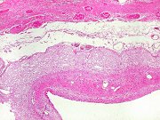
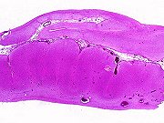
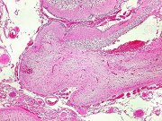
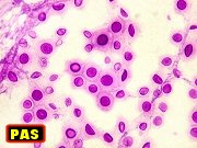
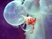
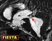
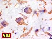
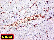
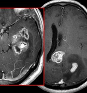
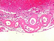
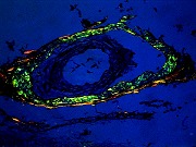
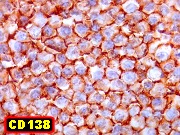
| M. 56 yr.
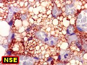
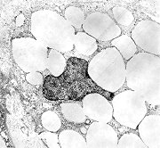
Granular cell tumor of neurohypophysis at III ventricle floor |
Same,
HE, special stains, IH |
|
|
|
|
|
|
 |
 |
 |
 |
| M.
6 yr. Adrenoleukodystrophy |
M.
11 yr. Adrenoleukodystrophy |
M.
10 yr. Adrenoleukodystrophy |
M.
7 yr. Metachromatic leukodystrophy |
| Leukodystrophies
- Illustrated linked text (in Portuguese) |
|
 |
 |
 |
 |
| F.
30 yr. Probable metachromatic leukodystrophy, adult form |
F.
56 yr. Probable metachromatic leukodystrophy, adult form |
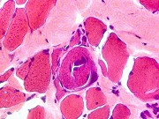
M.
5 yr. Leukodystrophy associated with congenital muscular dystrophy due
to merosin deficiency. Muscle biopsy |
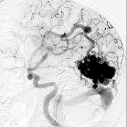
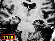
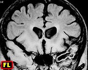
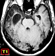
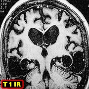
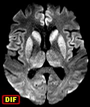
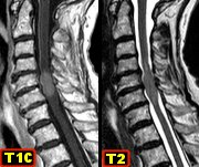
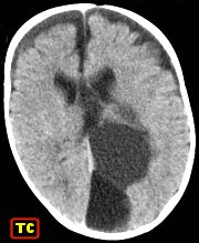
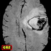
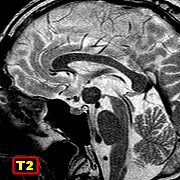
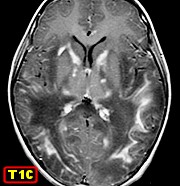
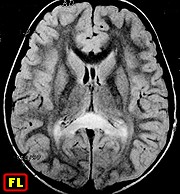
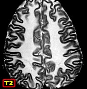
M.
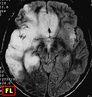
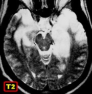
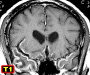
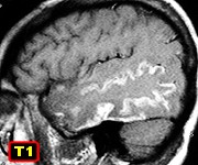
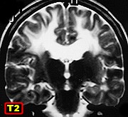
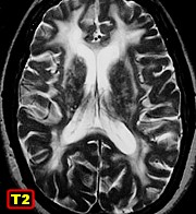
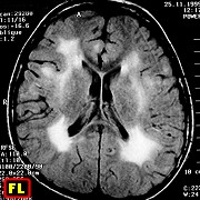
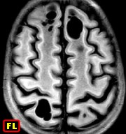
11 yr. van der Knaap's disease (megalencephalic
leukoencephalopathy with subcortical cysts). 11-year follow-up. Selection
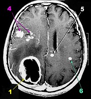
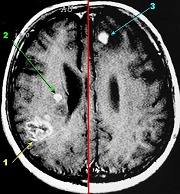
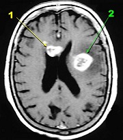
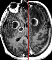
from 4 MRIs. |
|
|
|
Exams
in full |
|
|
|
|
| Neuro
Image Bank |
 |
 |
 |
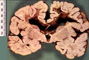 |
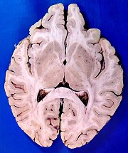
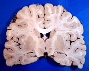
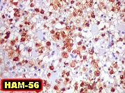
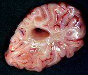
| Adrenoleukodystrophy.
Massive demyelination of frontal white matter sparing U fibers |
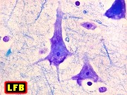
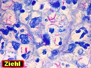
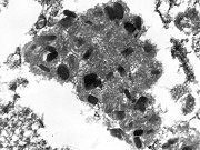
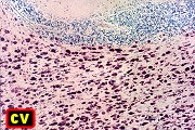
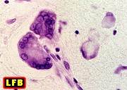
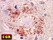
Metachromatic
leukodystrophy. Sulphatide storage in facial nerve nucleus in pons. |
Metachromatic
leukodystrophy. Sulphatide storage in macrophages in cerebellar white matter |
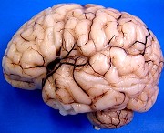
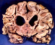
Sudanophilic
leukodystrophy. Cavitating demyelination of frontal white matter and ventricular
dilatation |
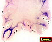
| Leukodystrophies
- Illustrated linked text (in Portuguese) |
|
 |
 |
 |
 |
| Sudanophilic
leukodystrophy. Demyelination of temporal white matter sparing U fibers |
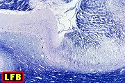
Krabbe's
globoid cell leukodystrophy - 3 cases. Demyelination
of occipital white matter sparing U fibers |
Krabbe's
globoid cell leukodystrophy. Globoid cells (macrophages) in cerebellar
white matter |
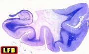
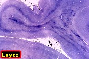
Pelizaeus
- Merzbacher's leukodystrophy. Tigroid appearance of cerebral white
matter |
|
|
|
|
 |
 |
 |
 |
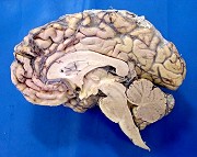
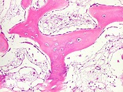
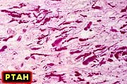
| Alexander's
leukodystrophy. Megalencephaly; softened friable hemispheric white matter |
Alexander's
leukodystrophy. Abundant Rosenthal fibers in lumbar spinal cord |
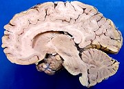
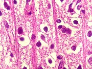
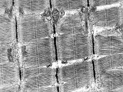
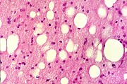
Canavan's
leukodystrophy. Spongy degeneration of hemispheric white matter |
M.
5 yr. Merosin deficient congenital muscular dystrophy and
leukodystrophy. Muscle biopsy. Brain MRI |
|
|
|
|
 |
 |
 |
 |
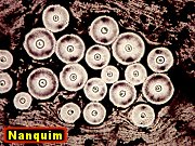
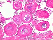
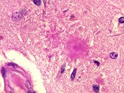
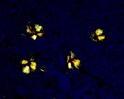
| F. 81 yr.
Senile plaques at hippocampus. HE, special stains |
Same, Congo
red and polarized light |
Same, IH |
Lhermitte-Duclos
disease (dysplastic gangliocytoma of cerebellum). Text |
|
|
|
|