| 30/5/2007 |
| New
Cases of Neuroimaging & Neuropathology: |
 |
 |
 |
 |
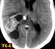
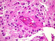
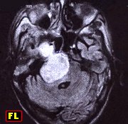
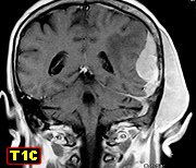
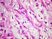
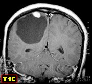
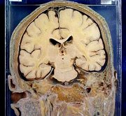
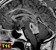
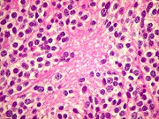
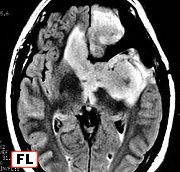
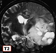
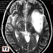
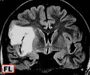
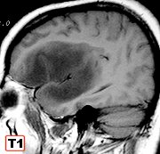
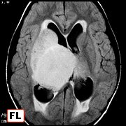
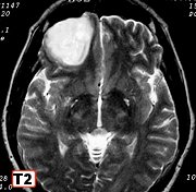
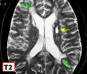
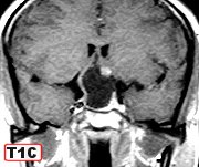
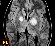
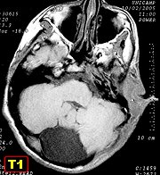
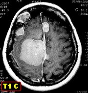
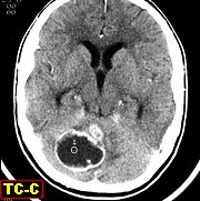
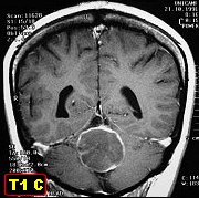
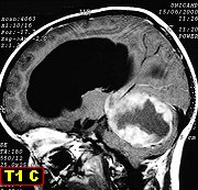
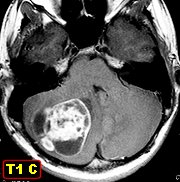
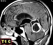
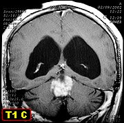
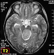
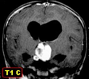
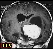
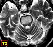
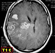
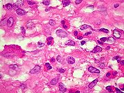
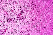
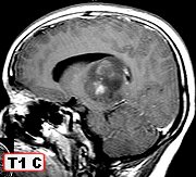
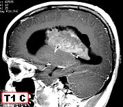
| F. 11 yr.
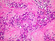
Diffuse astrocytoma of right thalamus with anaplastic foci |
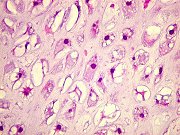
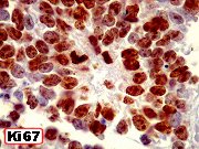
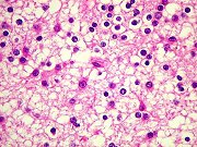
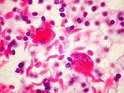
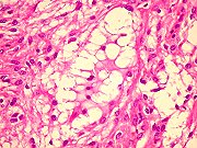
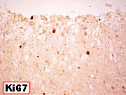
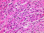
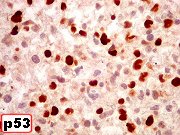
Same, HE,
IH. Pictured: high proportion of positive nuclei for p53 |
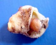
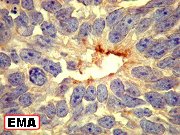
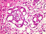
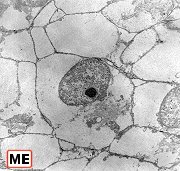
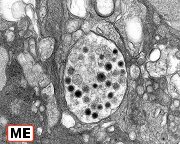
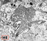
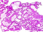
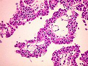
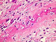
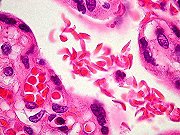
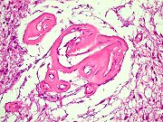
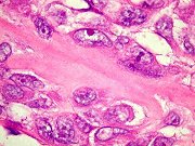
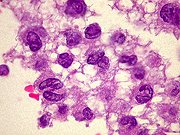
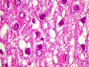
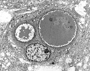
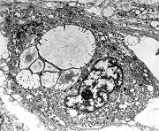
Secretory
meningioma. Electron microscopy. Pictured: pseudopsammomatous bodies |
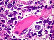
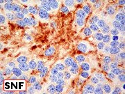
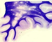
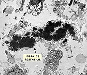
Pilocytic
astrocytoma of cerebellum rich in Rosenthal fibers. Electron microscopy |
|
|
|
|
 |
 |
 |
 |
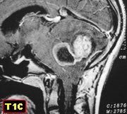
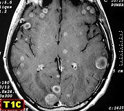
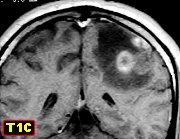
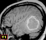
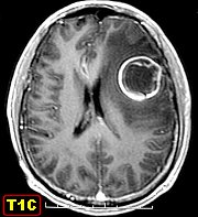
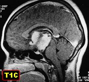
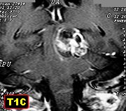
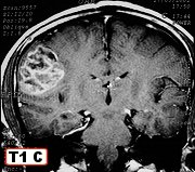
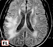
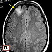
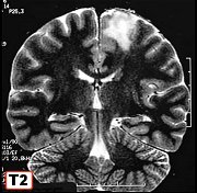
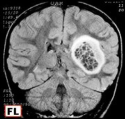
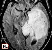
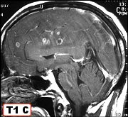
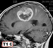
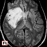
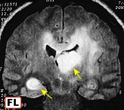
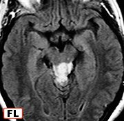
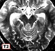
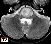
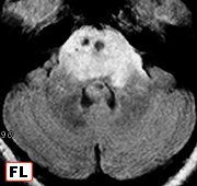
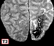
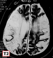
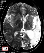
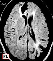
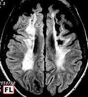
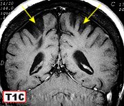
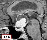
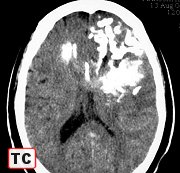
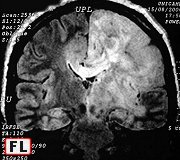
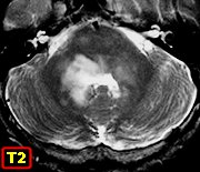
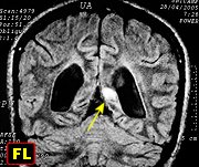
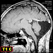
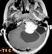
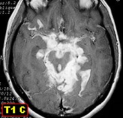
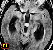
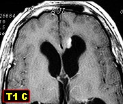
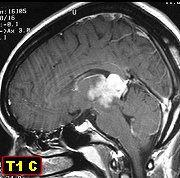
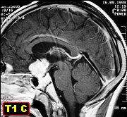
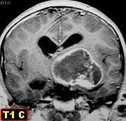
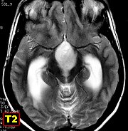
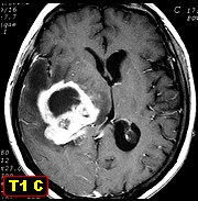
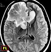
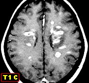
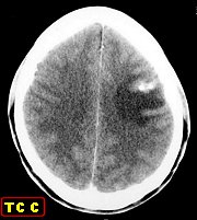
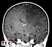
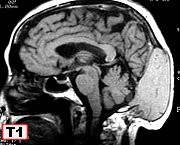
| F. 6 yr.
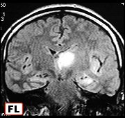
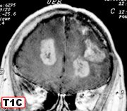
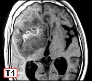
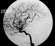
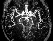
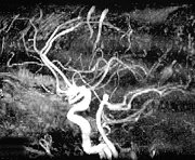
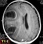
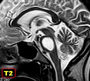
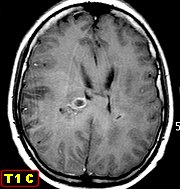
Gliomatosis cerebri (anaplastic diffuse astrocytoma of rapid growth). 1st
MRI |
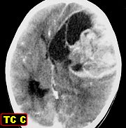
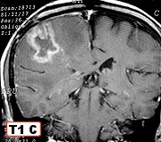
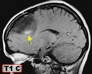
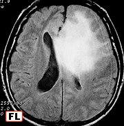
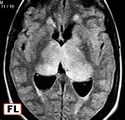
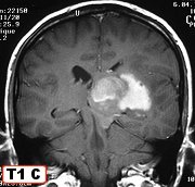
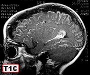
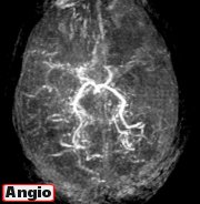
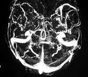
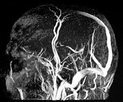
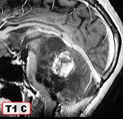
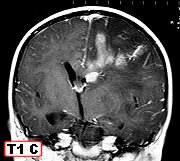
Same, after
40 days. Multiple foci of contrast enhancement |
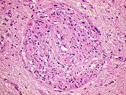
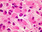
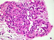
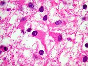
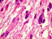
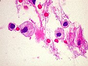
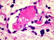
Same, stereotaxic
biopsy, HE, IH. Nuclear atypia, endothelial proliferation, thrombosis |
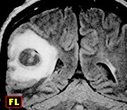
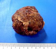
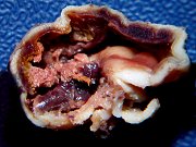
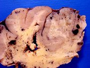
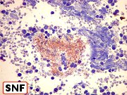
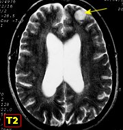
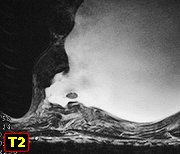
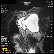
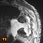
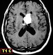
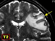
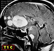
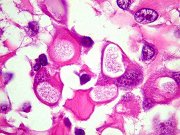
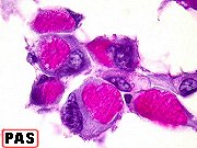
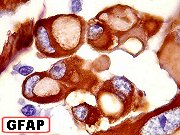
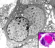
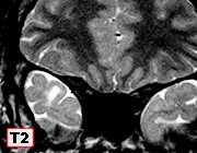
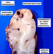
F. 14 yr.
Solid-cystic temporal
xanthoastrocytoma |
|
|
|
|
 |
 |
 |
 |
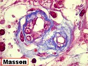
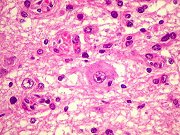
| Same, HE,
glycogen deposits in tumor cell cytoplasm |
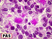
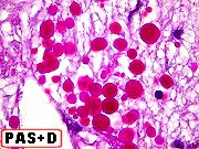
Same,
PAS |
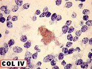
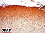
Same, IH |
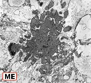
Same,
EM |
|
|
|
|
 |
 |
 |
 |
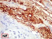
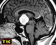
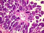
| F. 18 yr.
Central neurocytoma |
Same, HE,
IH. Pictured: astrocytic differentiation |
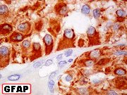
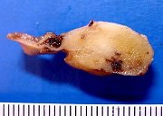
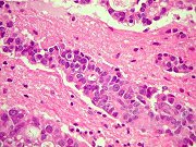
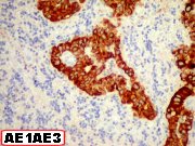
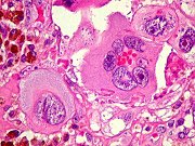
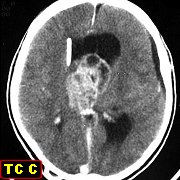
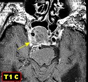
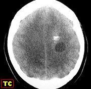
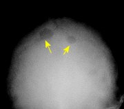
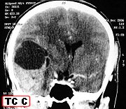
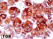
F. 75 yr.
Occipital metastasis of follicular carcinoma of thyroid. 4-year history |
Same, HE,
IH |
|
|
|
|
 |
 |
 |
 |
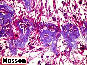
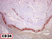
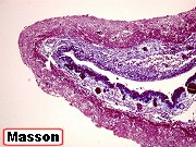
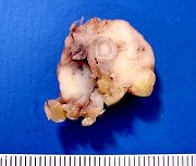
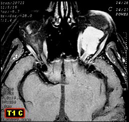
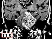
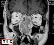
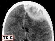
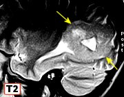
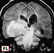
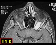
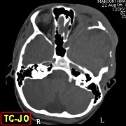
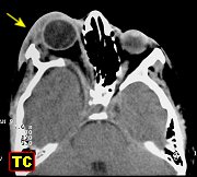
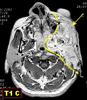
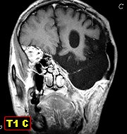
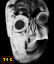
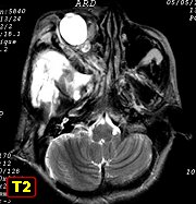
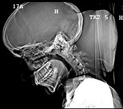
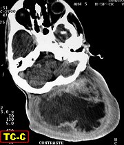
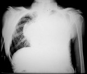
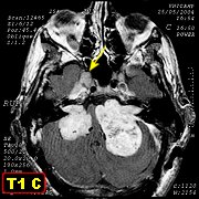
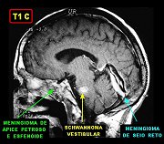
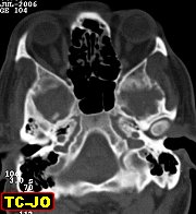
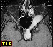
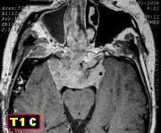
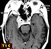
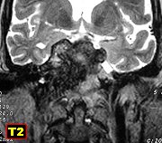
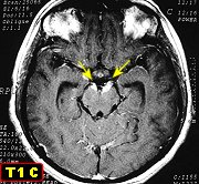
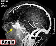
| Secretory
meningioma of sphenoid wing with hyperostosis. CT, MRI |
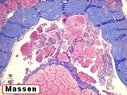
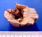
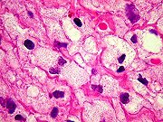
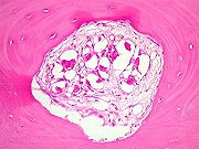
Same, intraosseous,
intracranial and intraorbitary components, HE |
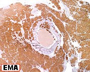
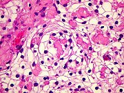
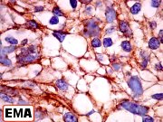
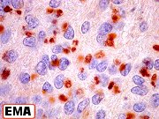
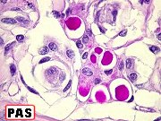
Same, PAS,
IH |
Same, EM |
|
|
|
|
 |
 |
 |
 |
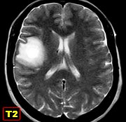
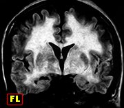
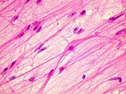
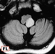
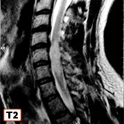
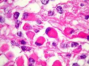
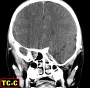
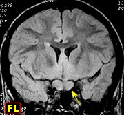
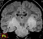
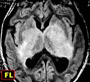
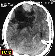
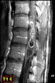
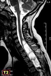
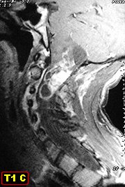
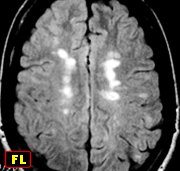
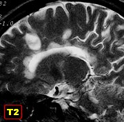
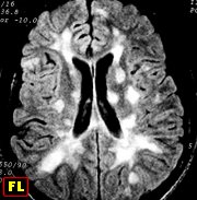
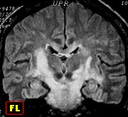
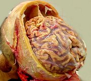
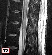
| M. 25 yr.
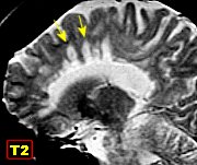
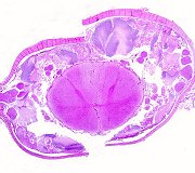
Systemic T/NK cell lymphoma masquerading as multiple sclerosis |
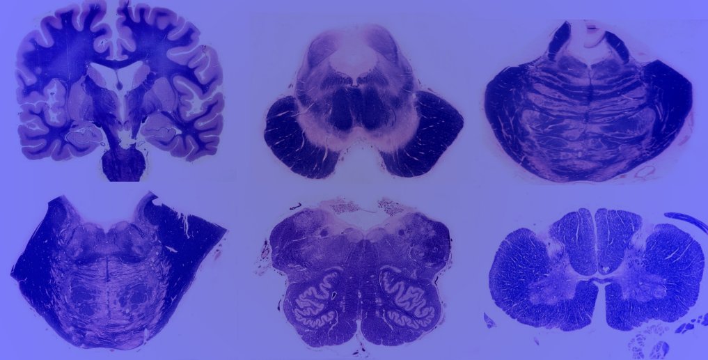
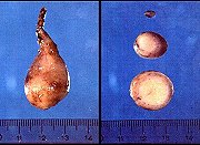
Same, postmortem
examination of brain and spinal cord |
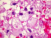
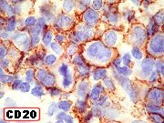
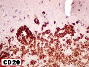
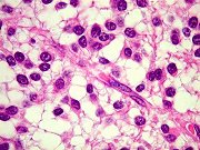
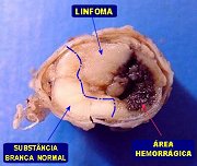
Same, brain,
HE - lymphoma |
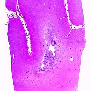
Same, spinal
cord, HE - lymphoma |
| Case
summary |
Texts: |
NK
cells |
T/NK
lymphomas |
 |
 |
 |
 |
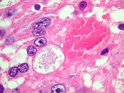
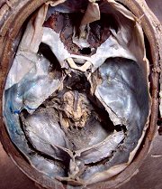
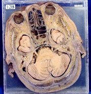
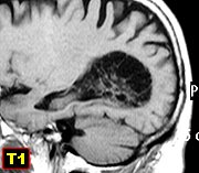
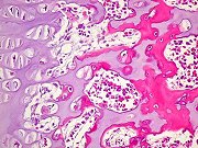
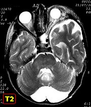
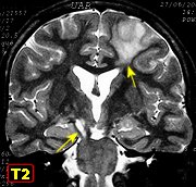
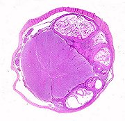
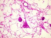
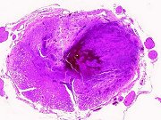
| Same, spinal
cord, HE - other findings. Pictured: cerebellar tonsil herniation down
to midthoracic cord |
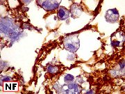
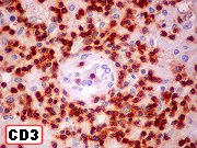
Same, spinal
cord, IH - lymphoma |
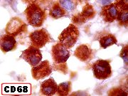
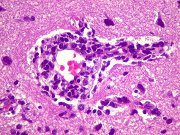
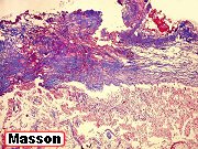
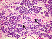
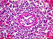
Same, other
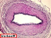
organs, HE. Pictured: lymphomatous infiltration of spleen arterioles |
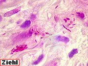
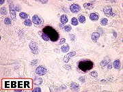
Same, endocardium,
IH. Pictured: nuclear positivity for Epstein-Barr virus encoded RNAs (EBER) |
|
|
|
|
 |
 |
 |
 |
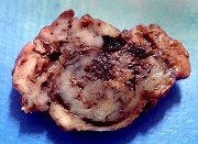
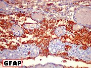
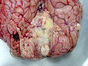
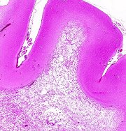
M. 15 yr.
Non-Taylor cortical dysplasia + incidental pleomorphic
xanthoastrocytoma |
Same, temporal
lobectomy, gross, HE |
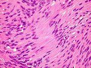
Same, dysplastic
cortex and white matter, HE |
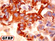
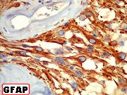
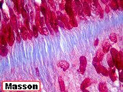
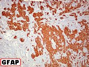
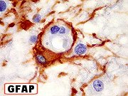
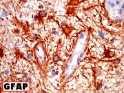
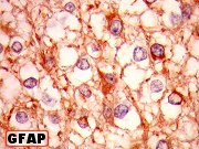
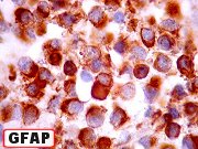
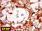
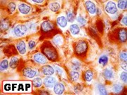
Same, GFAP.
Pictured: gliosis of molecular layer |
| Case
summary |
|
|
|
 |
 |
 |
|
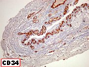
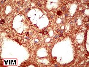
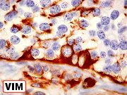
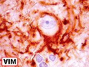
| Same, VIM |
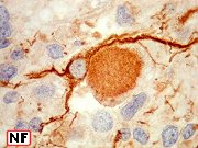
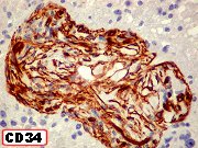
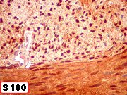
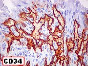
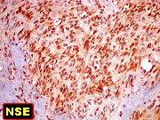
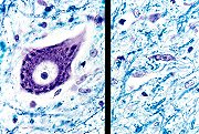
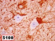
Same, S-100,
CD34, NSE. Pictured: abnormal neuron /astrocyte relationship |
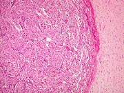
Same, intracortical
pleomorphic
xanthoastrocytoma,
(chance finding). HE, IH |
|
|
|
|
|
Neuro Image
Bank
»»» |
|
 |
|
|
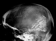
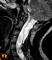
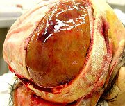
Massive
cerebellar herniation into spinal subarachnoid space down to midthoracic
level |
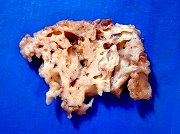
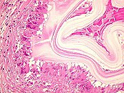
Corpora
amylacea |
|
|
|
|
|