| 7/11/2006 | |||
| New Cases of Neuroimaging & Neuropathology: | |||
 |
 |
 |
 |
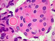
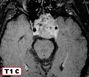
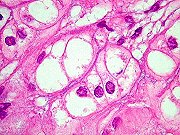
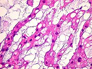
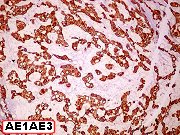
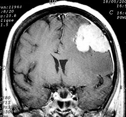
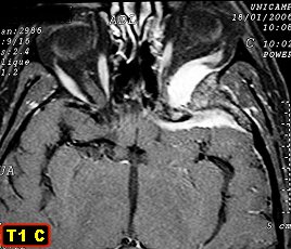
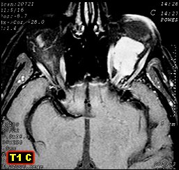
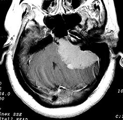
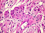
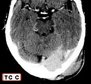
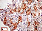
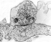
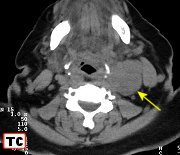
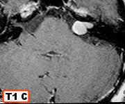
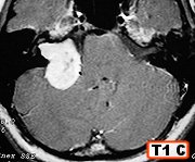
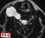
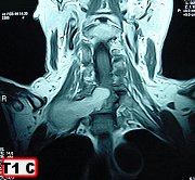
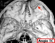
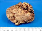
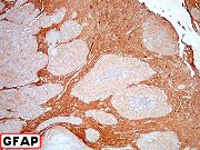
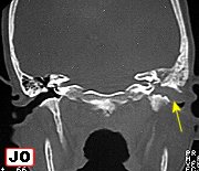
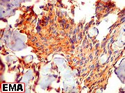
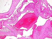
| Meningioma originating in middle ear | Same, HE, IH | Same, intracranial extension after 4 years. | Same, 2nd. specimen, HE, IH |
 |
 |
 |
|
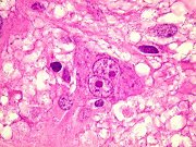
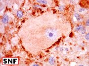
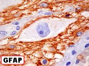
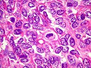
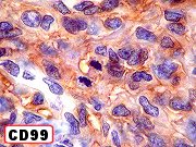
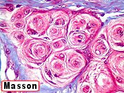
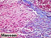
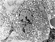
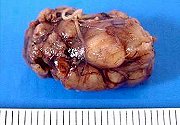
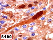
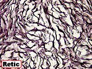
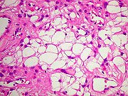
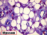
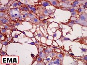
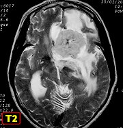
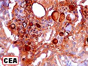
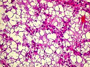
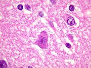
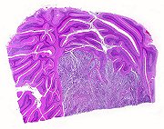
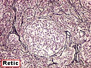
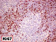
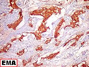
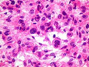
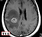
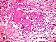
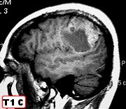
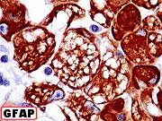
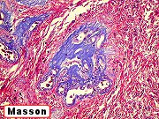
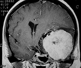
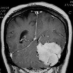
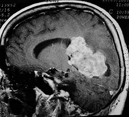
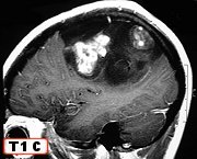
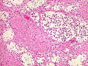
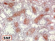
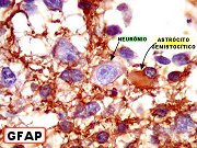
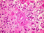
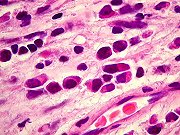
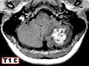
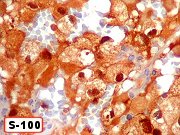
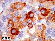
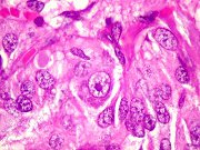
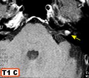
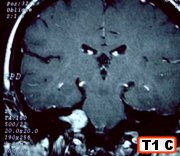
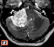
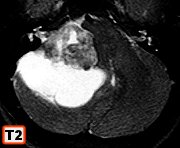
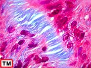
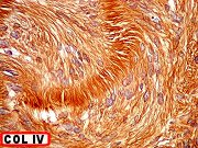
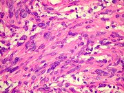
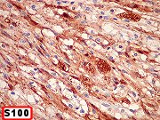
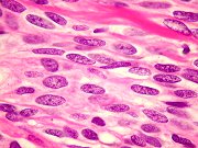
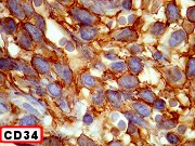
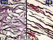
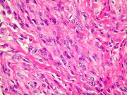
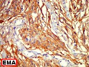
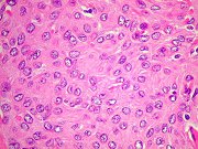
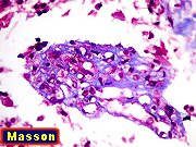
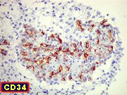
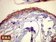
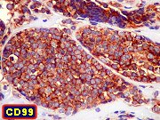
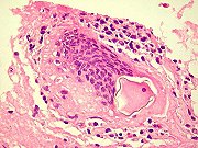
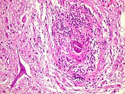
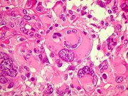
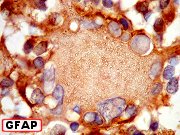
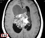
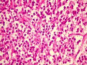
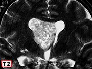
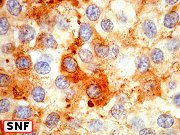
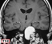
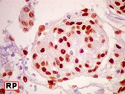
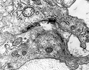
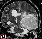
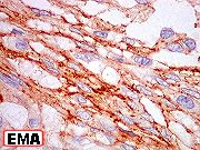
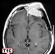
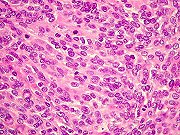
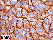
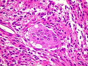
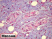
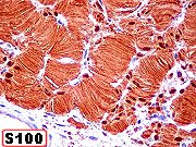
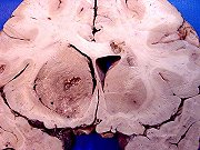
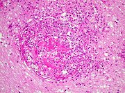
| Atypical meningioma | Same, HE, special stains | Same, IH. Text | |
 |
 |
 |
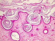 |
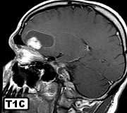
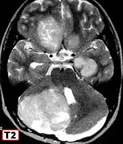
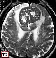
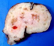
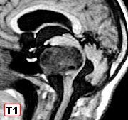
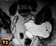
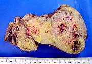
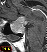
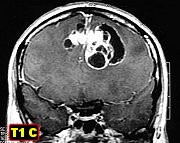
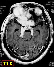
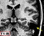
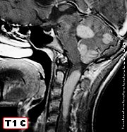
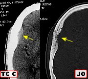
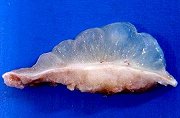
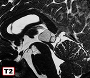
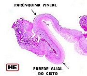
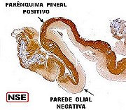
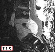
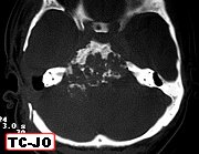
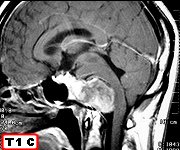
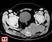
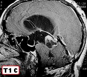
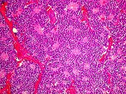
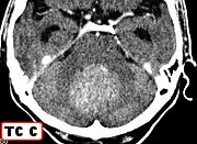
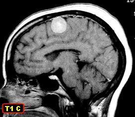
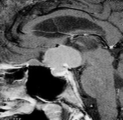
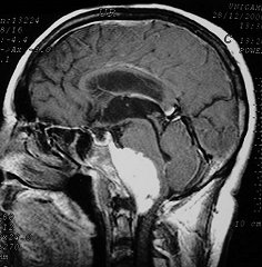
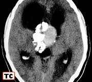
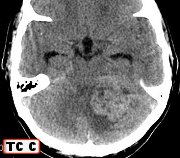
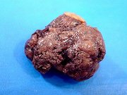
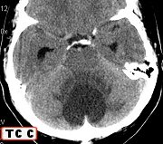
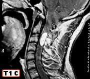
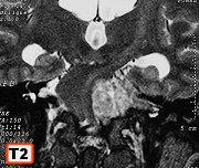
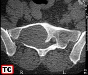
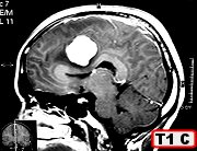
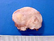
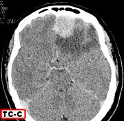
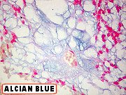
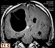
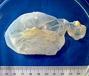
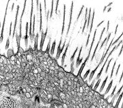
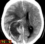
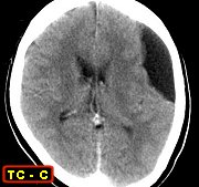
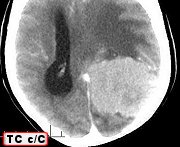
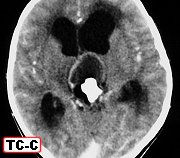
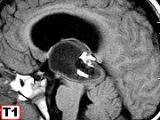
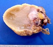
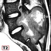
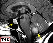
| Teratoma (dermoid cyst) of pineal - CT | Same, MRI | Same, macro specimen | Same, HE |
 |
 |
 |
 |
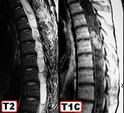
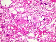
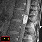
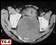
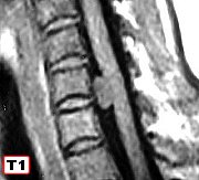
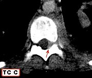
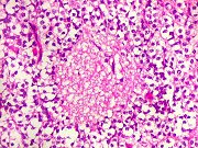
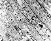
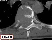
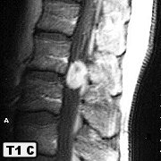
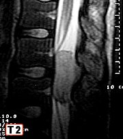
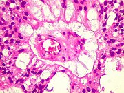
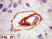
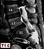
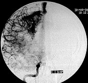
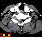
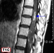
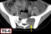
| Giant cell tumor of sacrum and lumbar vertebrae. | Same, HE. Text | Aneurysmal bone cyst of sacrum | Same, HE. Text |
 |
 |
||
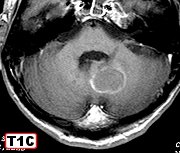
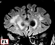
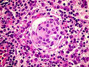
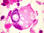
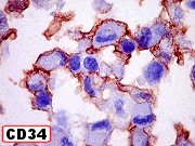
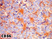
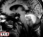
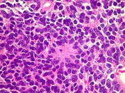
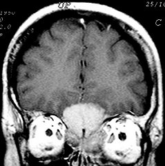
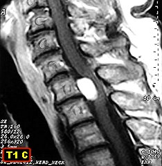
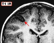
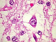
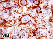
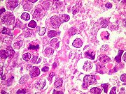
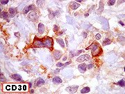
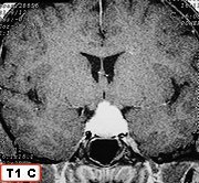
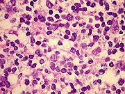
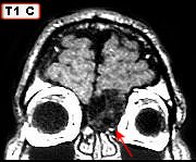
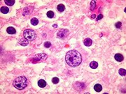
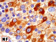
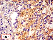
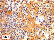
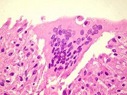
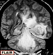
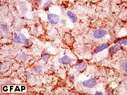
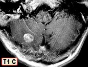
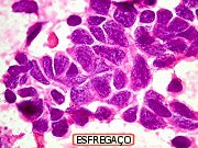
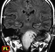
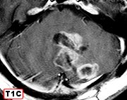
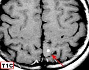
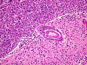
| B-cell lymphoma of cerebellum and pituitary | Same, HE, special stains, IH | ||
 |
 |
 |
|
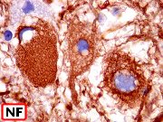
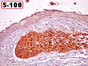
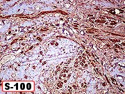
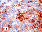
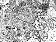
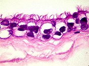
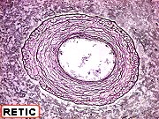
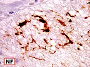
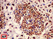
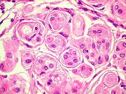
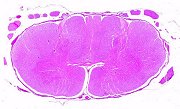
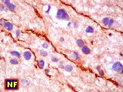
| Cutaneous plexiform neurofibroma. Macro, HE | Same, special stains | Same, IH | |
| Inflammations
Image Bank »»» |
 |
 |
 |
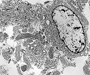
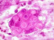
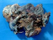
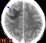
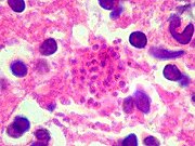
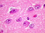
| Cerebral aspergillosis. Macro specimen | Same, HE | Same, Grocott | |
 |
 |
 |
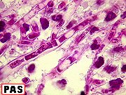 |
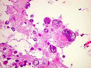
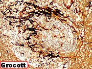
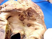
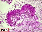
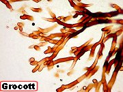
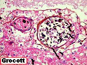
| Systemic aspergillosis. Macro specimens | Same, lung, HE, PAS | Same, lung, Grocott | Same, heart, HE, PAS, Grocott |
 |
 |
 |
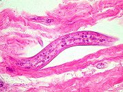 |
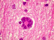
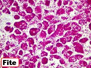
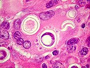
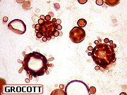
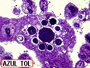
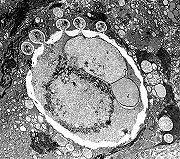
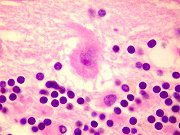
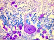
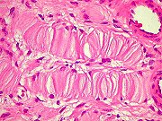
| Same, brain, HE+Grocott | Histoplasmosis and paracoccidioidomycosis of lung in AIDS. HE, Grocott | Intestinal strongyloidiasis in AIDS | |