| 14/6/2021 |
|
|
|
|
 |
 |
|
 |
 |
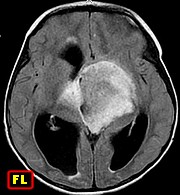
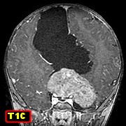
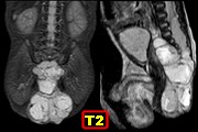
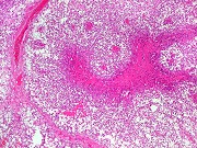
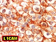
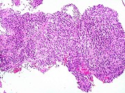
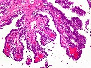
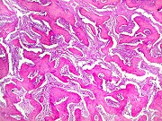
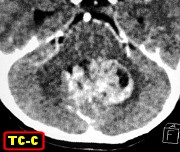
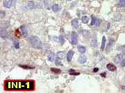
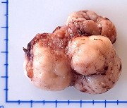
| M.
10 m 8 d. Atypical teratoid / rhabdoid tumor of posterior fossa. |
|
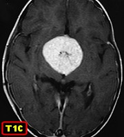
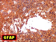
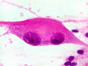
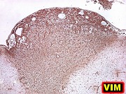
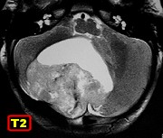
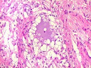
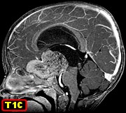
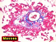
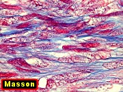
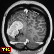
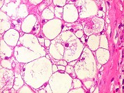
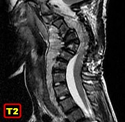
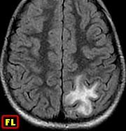
F.
7 yr 6 m Meningioangiomatosis in left parietal lobe |
| . |
.. |
. |
 |
 |
|
 |
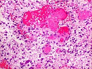 |
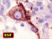
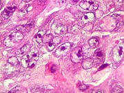
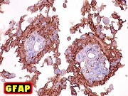
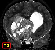
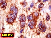
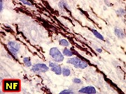
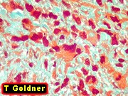
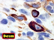
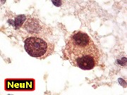
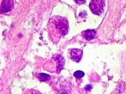
| F.
14 yr. Medullomyoblastoma followed by anaplastic cerebellar ganglioglioma
after 11 years |
|
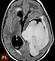
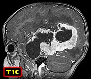
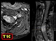
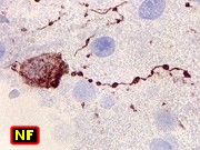
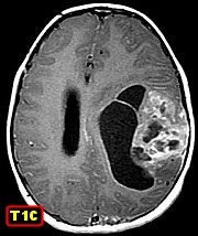
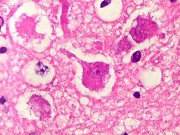
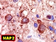
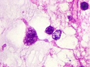
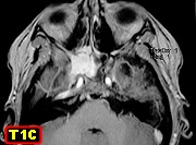
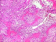
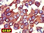
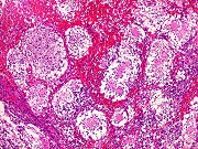
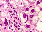
(Same
case) F. 24 yr. Anaplastic cerebellar ganglioglioma 11 years after
medullomyoblastoma (transformation or new tumor ?) |
|
|
|
|
|
 |
 |
|
 |
 |
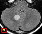
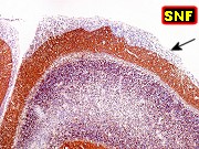
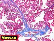
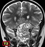
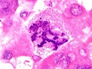
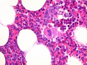
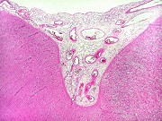
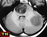
| F.
6 yr 11 m. Medulloblastoma with extensive nodularity in cerebellar vermis. |
|
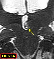
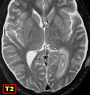
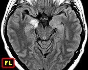
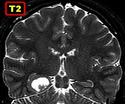
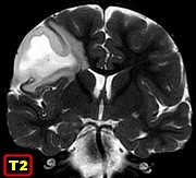
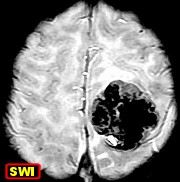
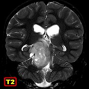
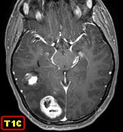
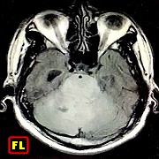
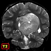
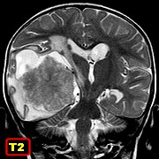
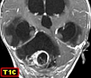
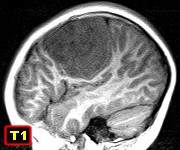
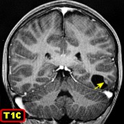
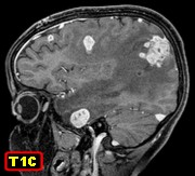
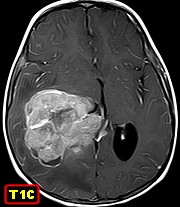
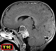
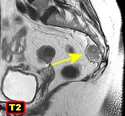
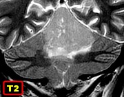
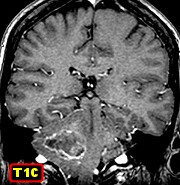
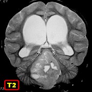
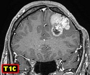
M.
7 yr 9 m. Diffuse midline glioma of pons with extension to left cerebellar
hemisphere |
|
|
|
|
|
 |
 |
|
 |
 |
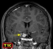
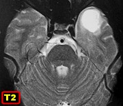
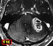
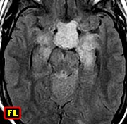
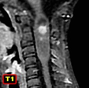
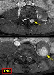
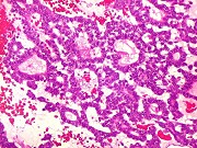
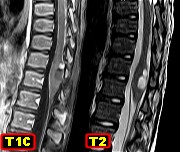
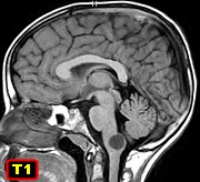
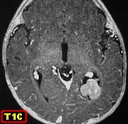
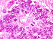
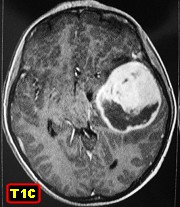
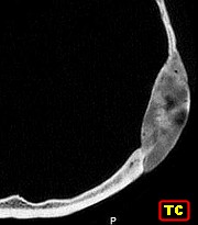
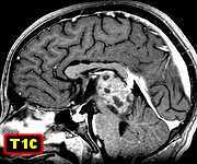
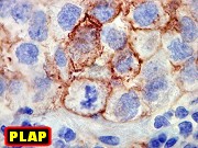
| M.
13 yr 6 m. Germinoma of pineal region filling III ventricle. |
|
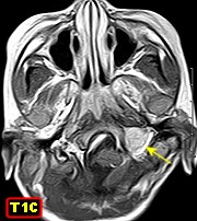
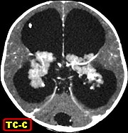
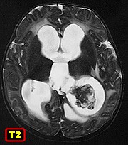
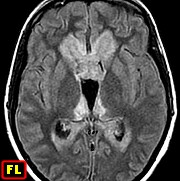
F.
15 yr 2 m. Germinoma of III ventricle spreading throughout the ventricular
system |
|
|
|
|
|
|
 |
|
 |
|
|
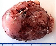
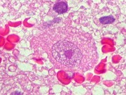
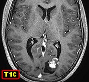
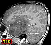
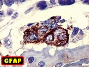
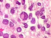
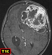
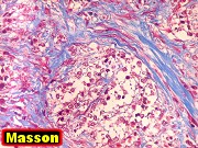
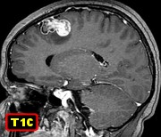
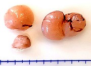
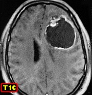
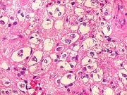
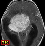
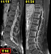
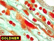
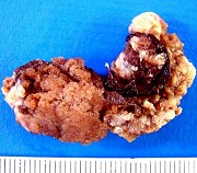
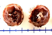
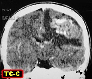
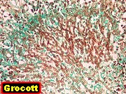
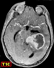
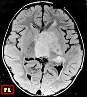
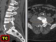
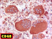
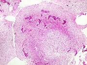
M.
27 yr 2 m. Primary sarcoma of cerebral cortex recurring after 8 years |
|