| 28/12/10 | |||
| New Cases of Neuroimaging & Neuropathology: | |||
 |
 |
 |
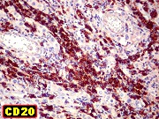 |
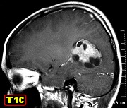
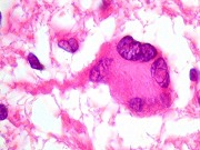
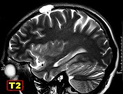
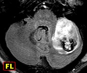
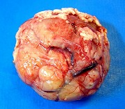
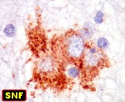
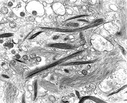
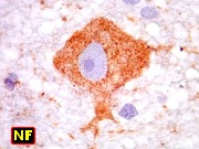
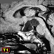
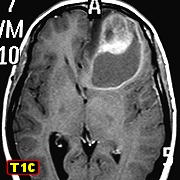
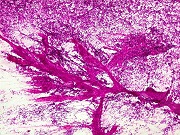
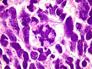
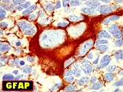
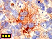
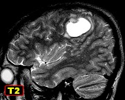
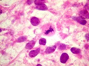
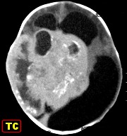
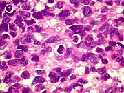
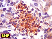
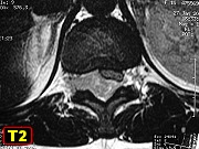
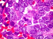
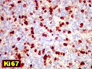
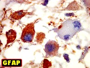
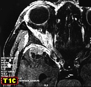
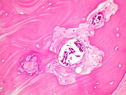
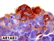
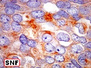
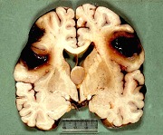
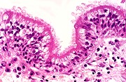
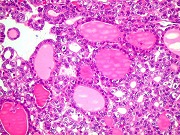
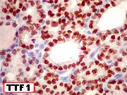
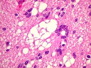
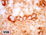
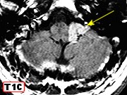
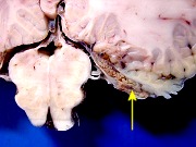
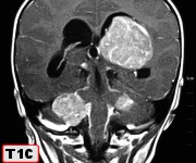
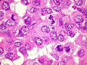
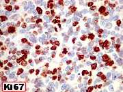
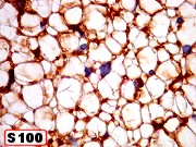
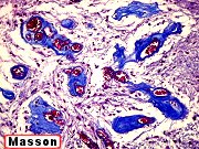
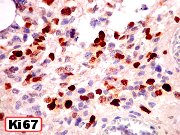
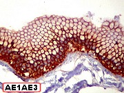
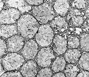
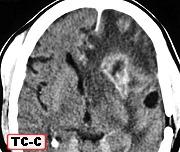
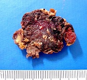
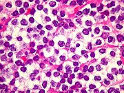
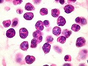
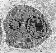
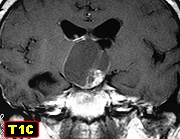
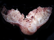
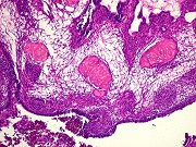
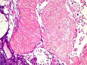
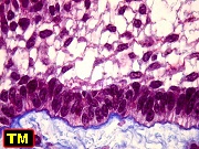
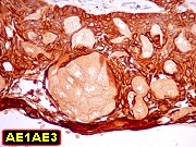
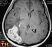
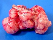
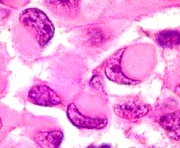
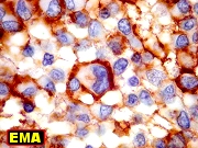
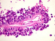
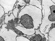
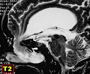
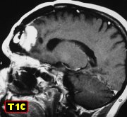
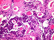
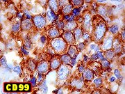
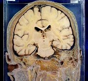
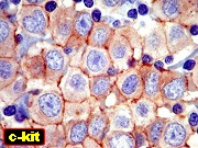
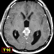
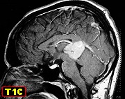
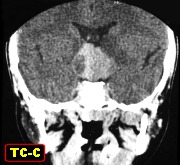
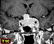
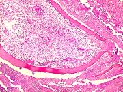
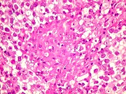
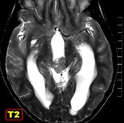
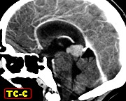
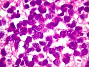
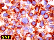
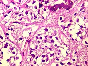
| M. 29 yr. Germinoma of pineal | Same, specimen, HE | Same, immunohistochemistry for PLAP, c-kit, b-HCG, AE1AE3. | Same, IH - CD20, CD3, vimentin, Ki-67. |
| Text : germinal tumors | Text : tumors of pineal parenchyma | Texts: PLAP, c-kit | |
 |
 |
 |
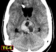 |
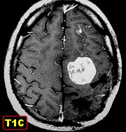
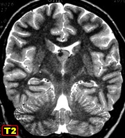
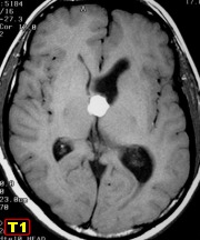
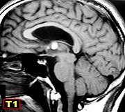
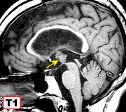
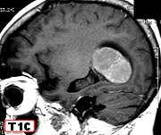
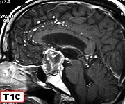
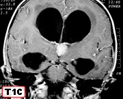
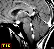
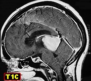
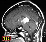
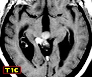
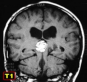
| M. 21 yr. Germinoma of pineal | M. 19 yr. Germinoma of pineal | M. 9 yr. Germinoma of pineal | M. 28 yr. Germinoma of pineal |
 |
 |
 |
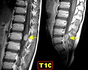 |
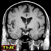
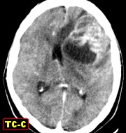
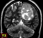
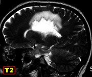
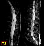
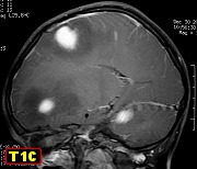
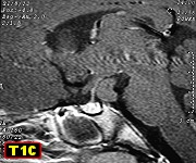
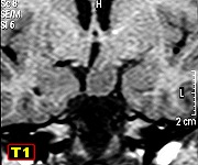
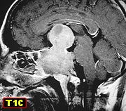
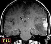
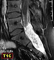
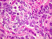
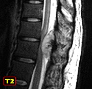
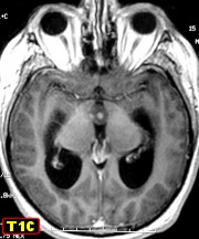
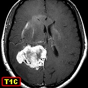
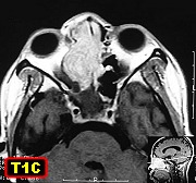
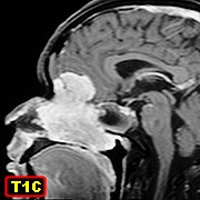
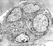
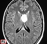
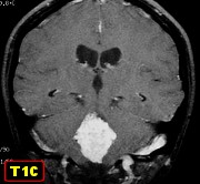
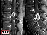
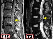
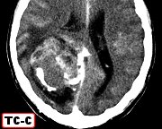
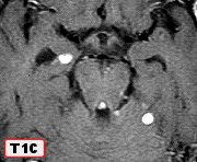
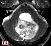
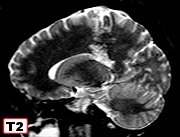
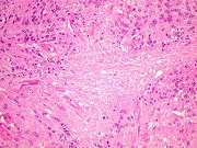
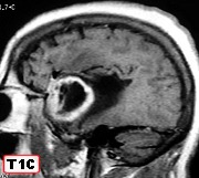
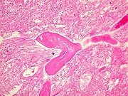
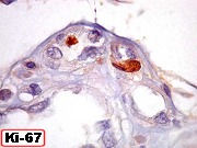
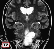
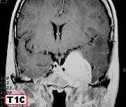
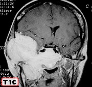
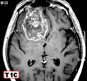
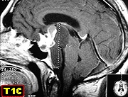
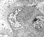
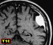
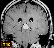
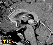
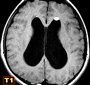
| F. 12 yr. Germinoma of hypothalamic region | M. 21 yr. Germinoma of hypothalamic region infiltrating dorsum sellae, cavernous sinuses and compressing optic chiasm | Same, HE, special stains, IH - c-kit, Ki-67 | M. 9 yr. Germinoma of pineal. Seeding of cranial subarachnoid space and lumbar nerve roots. Two-year follow-up. |
 |
 |
 |
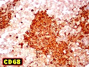 |
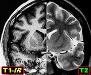
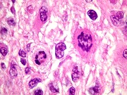
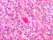
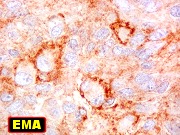
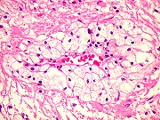
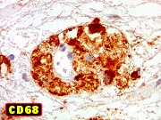
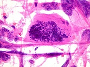
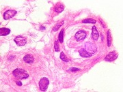
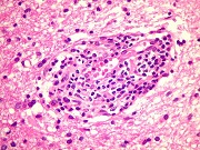
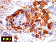
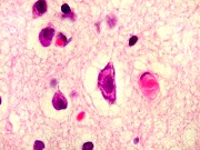
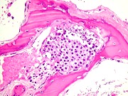
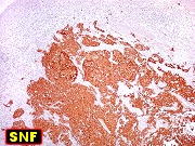
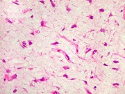
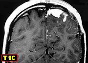
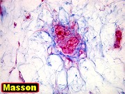
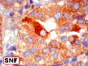
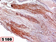
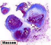
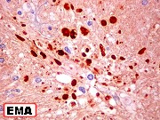
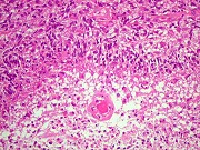
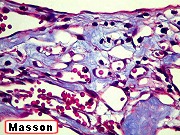
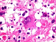
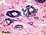
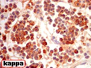
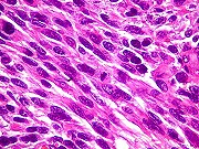
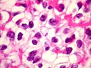
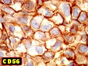
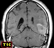
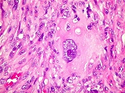
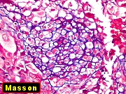
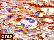
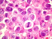
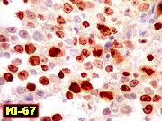
| F. 16 yr. Germinoma of hypothalamic region disseminating to IV ventricle | Same, HE | M. 17 yr. Germinoma of pineal with non caseating, epithelioid cell granulomas. HE | Same, IH - PLAP, c-kit, CD3, CD20, CD68 |
 |
 |
 |
 |
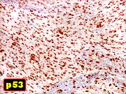
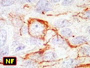
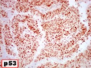
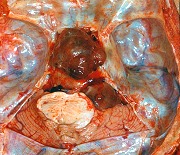
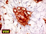
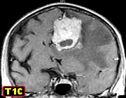
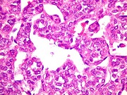
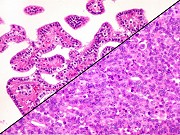
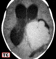
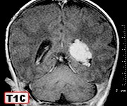
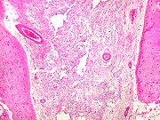
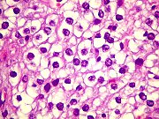
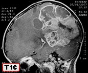
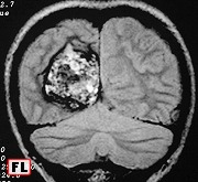
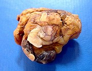
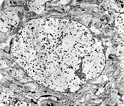
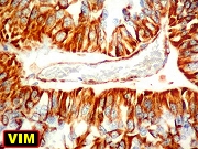
| Same, IH - VIM, AE1AE3, GFAP, NF, Ki-67, p53 | F. 24 yr. Pineocytoma. Histology for this case already available | F. 20 yr. Pineoblastoma. Histology for this case already available | M. 15 yr. Pineoblastoma |
 |
 |
 |
|
| F. 14 yr. Pineoblastoma | Same, HE, special stains | Same, IH | |
 |
 |
 |
 |
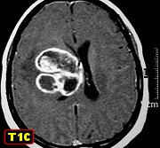
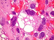
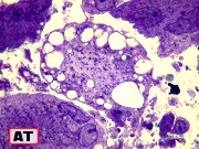
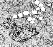
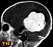
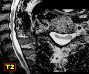
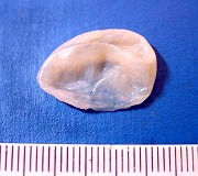
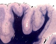
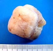
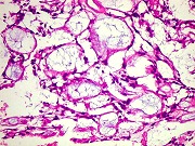
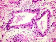
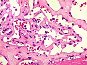
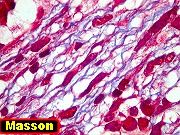
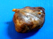
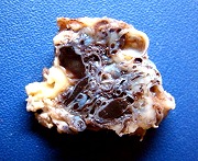
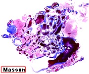
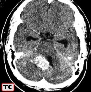
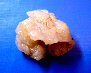
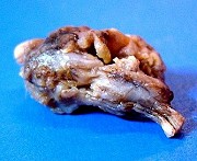
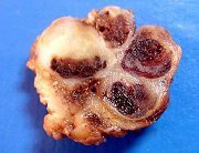
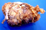
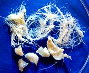
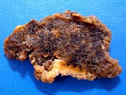
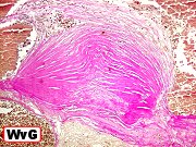
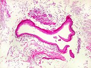
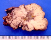
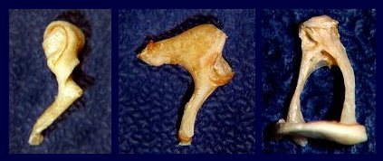
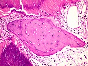
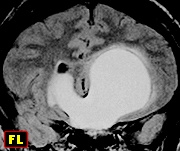
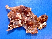
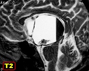
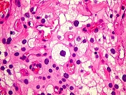
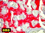
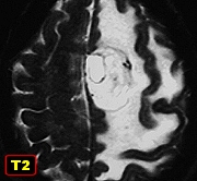
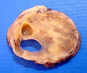
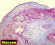
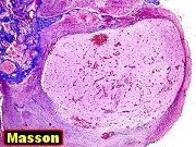
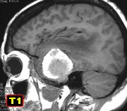
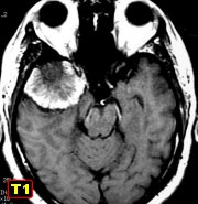
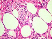
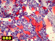
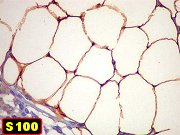
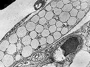
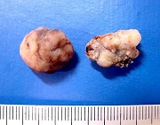
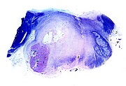
| F. 58 yr. Three-lobed cyst of pineal | Same, HE, IH | M. 13 yr. Teratoma of pineal. Free lipid droplets in lateral ventricles | |
 |
 |
 |
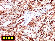 |
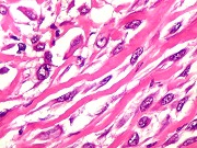
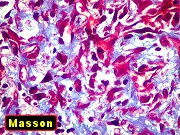
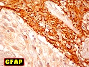
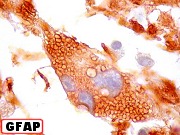
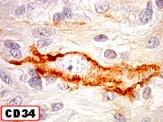
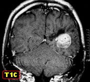
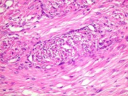
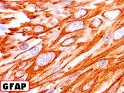
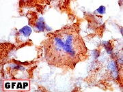
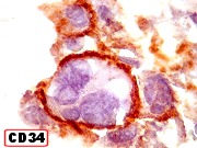
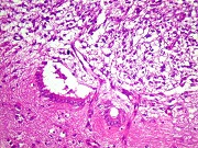
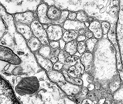
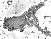
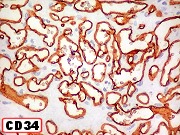
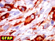
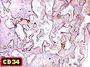
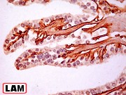
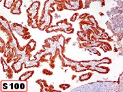
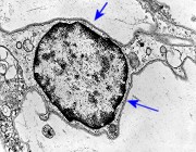
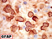
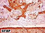
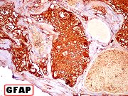
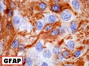
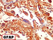
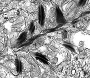
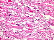
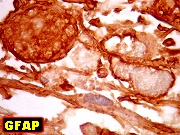
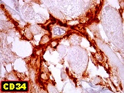
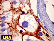
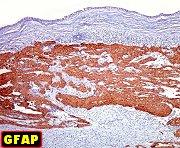
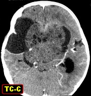
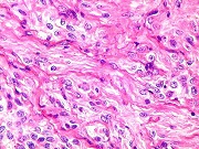
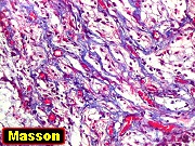
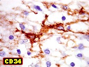
| M. 1 yr. Desmoplastic infantile astrocytoma | Same, HE, text | Same, special stains | Same, IH - GFAP, VIM, synaptophysin |
 |
»»» |
 |
 |
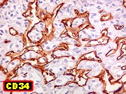
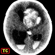
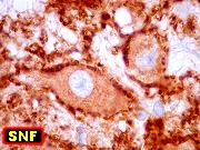
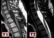
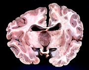
| Same, IH - CD34, 1A4, Ki-67, p53 | Pineal tumors | Brain stem gliomas | |